


Page 274 - Textbook of Pathology, 6th Edition
P. 274
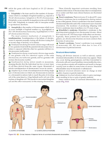
258 while the germ cells have haploid or 1N (23 chromo-     Three clinically important syndromes resulting from
           somes).                                             numerical aberrations of chromosomes due to nondisjunction
           1. Polyploidy is the term used for the number of chromo-  are as under and their main clinical features are illustrated
           somes which is a multiple of haploid number e.g. triploid or  in Fig. 10.3:
           3N (69 chromosomes), tetraploid or 4N (92 chromosomes).  Down’s syndrome. There is trisomy 21 in about 95% cases
           Polyploidy occurs normally in megakaryocytes and dividing  of Down’s syndrome due to nondisjunction during meiosis
           liver cells. Polyploidy in somatic cells of conceptus results  in one of the parents. Down’s syndrome is the most common
           in spontaneous abortions.                           chromosomal disorder and is the commonest cause of mental
                                                               retardation. The incidence of producing offspring with
           2. Aneuploidy is the number of chromosomes which is not  Down’s syndrome rises in mothers over 35 years of age.
     SECTION I
           an exact multiple of haploid number e.g. hypodiploid or  Klinefelter’s syndrome. Klinefelter’s syndrome is the
           2N-1 (45 chromosomes) monosomy, hyperdiploid or 2 N+1  most important example of sex chromosome trisomy. About
           (47 chromosomes) trisomy.                           80% cases have 47, XXY karyotype while others are mosaics.
              The most common mechanism of aneuploidy is       Typically, these patients have testicular dysgenesis. In
           nondisjunction. Nondisjunction is the failure of chromo-  general, sex chromosome trisomies are more common than
           somes to separate normally during cell division during first  trisomies of autosomes.
           or second stage of meiosis, or in mitosis.             Turner’s syndrome. Turner’s syndrome is an example
              Nondisjunction during first meiotic division stage will result  of monosomy (45, X0) most often due to loss of X
           in two gametes from both the parental chromosomes due to  chromosome in paternal meiosis.
           failure to separate while the other two gametes will have no
           chromosomes (nullisomic).
              Nondisjunction during second meiotic division stage results  Structural Abnormalities
           in one gamete with two identical copies of the same  During cell division (meiosis as well as mitosis), certain
           chromosome, one nullisomic gamete, and two gametes with  structural abnormalities of chromosomes may appear. These
           normal chromosome number.                           may occur during gametogenesis and then transmitted to
              Nondisjunction during mitosis results in mosaicism,  all somatic cells and cause hereditary transmissible disorders,
           meaning thereby that the individual has two or more types  or may produce somatic cell mutations and result in changes
           of cell lines derived from the same zygote. Mosaicism of  varying from no effect to some forms of cancers. Structural
           mitotic nondisjunction of chromosomes occurs in cancers.  abnormalities may be balanced or unbalanced.
              Anaphase lag is a form of nondisjunction involving single  Balanced structural alteration means no change in total
           pair of chromosomes in which one chromosome in meiosis  number of genes or genetic material.
     General Pathology and Basic Techniques
           or a chromatid in mitosis fails to reach the pole of dividing  Unbalanced structural alteration refers to gene rearrange-
           cell at the same time (i.e. it lags behind) and is left out of the  ment resulting in loss or gain of genetic material.
           nucleus of daughter cell. This results in one normal daughter  Some common forms of structural abnormalities are as
           cell and the other monosomic for the missing chromosome.  under (Fig. 10.4):


           Figure 10.3  Clinical features of important forms of numerical chromosomal abnormalities.

