


Page 28 - Textbook of Pathology, 6th Edition
P. 28
12
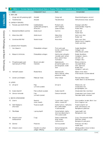
TABLE 2.1: Common Special (Histochemical) Stains in Surgical Pathology (in Alphabetic Order of Constituents).
Stain Component/Tissue Dyes Interpretation
A. AMYLOID
1. Congo red with polarising light Amyloid Congo red Green-birefringence: amyloid
2. Toluidine blue Amyloid Toluidine blue Orthochromatic blue: amyloid
B. CARBOHYDRATES
3. Periodic acid-Schiff (PAS) Carbohydrates Periodic acid, Glycogen and other
SECTION I
(particularly glycogen), Schiff reagent carbohydrates: magenta
all mucins (basic fuchsin) Nuclei: blue
4. Mucicarmine/Best’s carmine Acidic mucin Carmine Mucin: red
Nuclei: blue
5. Alcian blue (AB) Acidic mucin Alcian blue Acid mucin: blue
(at pH 2.5) Nuclei: red
6. Combined AB-PAS Neutral mucin Alcian blue Acid mucin: blue
Neutral mucin: magenta
Nuclei: pale blue
C. CONNECTIVE TISSUES
7. Van Gieson’s Extracellular collagen Picric acid, acid Nuclei: blue/black
fuchsin, celestin blue- Collagen: red
haemalum Other tissues: yellow
8. Masson’s trichrome Extracellular collagen Acid fuchsin, phospho- Nuclei: blue/black
molybdic acid, methyl Cytoplasm, muscle,
blue, celestin blue- red cells: red
haemalum Collagen: blue
9. Phosphotungstic acid- Muscle and glial Haematoxylin, Muscle striations,
haematoxylin (PTAH) filaments phosphotungstic acid, neuroglial fibres,
permanganate, oxalic fibrin: dark blue
acid Nuclei: blue
Cytoplasm: pale pink
10. Verhoeff’s elastic Elastic fibres Haematoxylin, Elastic fibres: black
General Pathology and Basic Techniques
Ferric chloride, iodine, Other tissues: counter-stained
potassium iodide
11. Gordon and Sweet’s Reticular fibres Silver nitrate Reticular fibres: black
Nuclei: black or counterstained
D. LIPIDS
12. Oil red O Fats Oil red O Mineral oils: red
(unfixed cryostat) Unsaturated fats,
phospholipids: pink
13. Sudan black B Fats (unfixed cryostat) Sudan black B Unsaturated fats: blue black
14. Osmium tetroxide Fats Osmium tetroxide Unsaturated lipids: brown black
(unfixed cryostat) Saturated lipids: unstained
E. MICRO-ORGANISMS
15. Gram’s Bacteria Crystal violet, Lugol’s Gram-positive, keratin, fibrin: blue
(cocci, bacilli) iodine, neutral red Gram-negative: red
16. Ziehl-Neelsen’s Tubercle bacilli Carbol fuchsin, methylene Tubercle bacilli, hair
(Acid-fast) blue (differentiate shaft, actinomyces: red
in acid-alcohol) Background: pale blue
17. Fite-Wade Leprosy bacilli Carbol fuchsin, methy- Lepra bacilli: red
lene blue (decolorise in Background: blue
10% sulfuric acid)
18. Grocott’s silver Fungi Sodium tetraborate, Fungi, Pneumocystis: black
methanamine silver nitrate, Red cells: yellow
methanamine Background: pale green
19. Giemsa Parasites Giemsa powder Protozoa: dark blue
Nuclei: blue
20. Shikata’s orcein Hepatitis B surface Acid permanganate, HBsAg positive: brown to black
antigen (HBsAg) orcein, tetrazine Background: yellow
Contd...

