


Page 30 - Textbook of Pathology, 6th Edition
P. 30
14                                                      Filters. A variety of filters are used between the source of
                                                               light and objective: first, heat absorbing filter; second, red-light
                                                               stop filter; and third exciter filter to allow the passage of light
                                                               of only the desired wavelength. On passing through the
                                                               specimen, light of both exciting and fluorescence wavelength
                                                               collects. Exciter light is removed by another filter called barrier
                                                               filter between the objective and the observer to protect the
                                                               observer’s eyes so that only fluorescent light reaches the eyes
                                                               of observer.
     SECTION I
                                                               Condenser. Dark-ground condenser is used in fluorescence
                                                               microscope so that no direct light falls into the object and
                                                               instead gives dark contrast background to the fluorescence.

                                                               TECHNIQUES.  There are two types of fluorescence
                                                               techniques both of which are performed on cryostat sections
                                                               of fresh unfixed tissue: direct and indirect.
                                                                   In the direct technique, first introduced by Coons (1941)
                                                               who did the original work on immunofluorescence, antibody
                                                               against antigen is directly conjugated with the fluorochrome
                                                               and then examined under fluorescence microscope.
                                                                   In the indirect technique, also called sandwich technique,
                                                               there is interaction between tissue antigen and specific anti-
                                                               body, followed by a step of washing and then addition of
                                                               fluorochrome for completion of reaction. Indirect
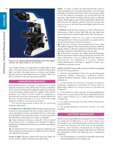
           Figure 2.5  Binocular light microscope (Model E 400, Nikon, Japan).
           Courtesy: Towa Optics (India) Pvt. Ltd., New Delhi.  immunofluorescence technique is applied to detect auto-
                                                               antibodies in patient’s serum.
           rays of light vibrate in a single plane at right angle to each  APPLICATIONS. Immunofluorescence methods are applied
           other. Two discs made up of prism are placed in the path of  for the following purposes:
           light, one below the object known as polariser and another  1. Detection of autoantibodies in the serum e.g. smooth muscle
           placed in the body tube which is known as analyser. The lower  antibodies (SMA), antinuclear antibodies (ANA),
     General Pathology and Basic Techniques
           disc is rotated to make the light plane polarised. .
                                                               antimitochondrial antibody (AMA), thyroid microsomal
                                                               antibody etc.
                     IMMUNOFLUORESCENCE
                                                               2. In renal diseases for detection of deposits of immuno-
           Immunofluorescence technique is employed to localise  globulins, complement and fibrin in various types of
           antigenic molecules on the cells by microscopic examination.  glomerular diseases by frozen section as discussed in
           This is done by using specific antibody against the antigenic  Chapter 22.
           molecule forming antigen-antibody complex at the specific  3. In skin diseases to detect deposits of immunoglobulin by
           antigenic site which is made visible by employing a  frozen section, particularly at the dermo-epidermal junction
           fluorochrome which has the property to absorb radiation in  and in upper dermis e.g. in various bullous dermatosis
           the form of ultraviolet light so as to be within the visible  (Chapter 26).
           spectrum of light in microscopic examination.       4. For study of mononuclear cell surface markers using mono-
              The immunofluorescent method has the following   clonal antibodies.
           essential components:
                                                               5. For specific diagnosis of  infective disorders e.g. viral
           FLUORESCENCE MICROSCOPE. Fluorescence microscopy    hepatitis.
           is based on the principle that the exciting radiation from
           ultraviolet light of shorter wavelength (360 nm) or blue light
           (wavelength 400 nm) causes fluorescence of certain substances  ELECTRON MICROSCOPY
           and thereafter re-emits light of a longer wavelength.  Electron microscope (EM) first developed in 1930s in Germany
              Some substances fluoresce naturally; this is termed
           primary fluorescence or autofluorescence  though UV light is  has undergone modifications so as to add extensive new
                                                               knowledge to our understanding the structure and function
           required for visualising them better e.g. vitamin A, porphyrin,  of normal and diseased cells at the level of cell organelles.
           chlorophyll.                                        However, more recently, widespread use of diagnostic
              Secondary fluorescence is more commonly employed and
           is the production of fluorescence on addition of dyes or chemi-  immunohistochemistry in surgical pathology has restricted
                                                               the application of EM to the following areas of diagnostic
           cals called fluorochromes.
                                                               pathology:
           Source of light. Mercury vapour and xenon gas lamps are  1. In renal pathology in conjunction with light microscopy
           used as source of light for fluorescence microscopy.   and immunofluorescence (Chapter 22).

