


Page 32 - Textbook of Pathology, 6th Edition
P. 32
16 membranous staining for leucocyte common antigen (LCA), predict the biologic behaviour of the tumour. These include:
nuclear staining for oestrogen-progesterone receptors (ER- proto-oncogenes (e.g. HER-2/neu overexpression in
PR), cytoplasmic staining for smooth muscle actin (SMA) etc. carcinoma breast), tumour suppressor genes or antioncogenes
IHC stains cannot be applied to distinguish between (e.g. Rb gene, p53), growth factor receptors (e.g. epidermal
neoplastic and non-neoplastic lesions, or between benign and growth factor receptor or EGFR), and tumour cell proliferation
malignant tumours. These distinctions have to be done by markers (e.g. Ki67, proliferation cell nuclear antigen PCNA).
traditional methods in surgical pathology. Analysis of tumours by these methods is a significant
improvement in management over the conventional
Major Applications of IHC prognostic considerations by clinical staging and histologic
SECTION I
grading.
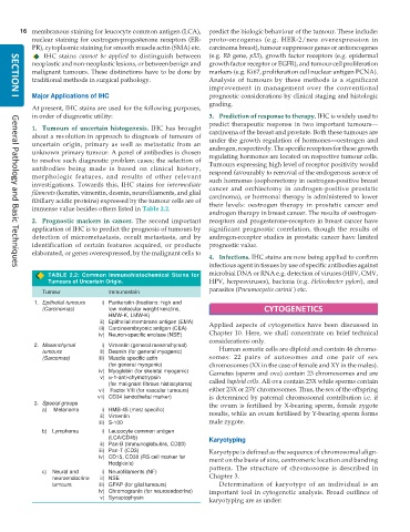
At present, IHC stains are used for the following purposes,
in order of diagnostic utility: 3. Prediction of response to therapy. IHC is widely used to
predict therapeutic response in two important tumours—
1. Tumours of uncertain histogenesis. IHC has brought carcinoma of the breast and prostate. Both these tumours are
about a revolution in approach to diagnosis of tumours of under the growth regulation of hormones—oestrogen and
uncertain origin, primary as well as metastatic from an androgen, respectively. The specific receptors for these growth
unknown primary tumour. A panel of antibodies is chosen regulating hormones are located on respective tumour cells.
to resolve such diagnostic problem cases; the selection of Tumours expressing high level of receptor positivity would
antibodies being made is based on clinical history, respond favourably to removal of the endogenous source of
morphologic features, and results of other relevant such hormones (oophorectomy in oestrogen-positive breast
investigations. Towards this, IHC stains for intermediate cancer and orchiectomy in androgen-positive prostatic
filaments (keratin, vimentin, desmin, neurofilaments, and glial carcinoma), or hormonal therapy is administered to lower
fibillary acidic proteins) expressed by the tumour cells are of their levels: oestrogen therapy in prostatic cancer and
immense value besides others listed in Table 2.2.
androgen therapy in breast cancer. The results of oestrogen-
2. Prognostic markers in cancer. The second important receptors and progesterone-receptors in breast cancer have
application of IHC is to predict the prognosis of tumours by significant prognostic correlation, though the results of
detection of micrometastasis, occult metastasis, and by androgen-receptor studies in prostatic cancer have limited
identification of certain features acquired, or products prognostic value.
elaborated, or genes overexpressed, by the malignant cells to
4. Infections. IHC stains are now being applied to confirm
infectious agent in tissues by use of specific antibodies against
General Pathology and Basic Techniques
TABLE 2.2: Common Immunohistochemical Stains for microbial DNA or RNA e.g. detection of viruses (HBV, CMV,
Tumours of Uncertain Origin. HPV, herpesviruses), bacteria (e.g. Helicobacter pylori), and
parasites (Pneumocystis carinii ) etc.
Tumour Immunostain
1. Epithelial tumours i) Pankeratin (fractions: high and
(Carcinomas) low molecular weight keratins, CYTOGENETICS
HMW-K, LMW-K)
ii) Epithelial membrane antigen (EMA) Applied aspects of cytogenetics have been discussed in
iii) Carcinoembryonic antigen (CEA)
iv) Neuron-specific enolase (NSE) Chapter 10. Here, we shall concentrate on brief technical
considerations only.
2. Mesenchymal i) Vimentin (general mesenchymal) Human somatic cells are diploid and contain 46 chromo-
tumours ii) Desmin (for general myogenic)
(Sarcomas) iii) Muscle specific actin somes: 22 pairs of autosomes and one pair of sex
(for general myogenic) chromosomes (XX in the case of female and XY in the males).
iv) Myoglobin (for skeletal myogenic) Gametes (sperm and ova) contain 23 chromosomes and are
v) α-1-anti-chymotrypsin called haploid cells. All ova contain 23X while sperms contain
(for malignant fibrous histiocytoma)
vi) Factor VIII (for vascular tumours) either 23X or 23Y chromosomes. Thus, the sex of the offspring
vii) CD34 (endothelial marker) is determined by paternal chromosomal contribution i.e. if
3. Special groups the ovum is fertilised by X-bearing sperm, female zygote
a) Melanoma i) HMB-45 (most specific) results, while an ovum fertilised by Y-bearing sperm forms
ii) Vimentin
iii) S-100 male zygote.
b) Lymphoma i) Leucocyte common antigen
(LCA/CD45) Karyotyping
ii) Pan-B (Immunoglobulins, CD20)
iii) Pan-T (CD3) Karyotype is defined as the sequence of chromosomal align-
iv) CD15, CD30 (RS cell marker for ment on the basis of size, centromeric location and banding
Hodgkin’s)
c) Neural and i) Neurofilaments (NF) pattern. The structure of chromosome is described in
neuroendocrine ii) NSE Chapter 3.
tumours iii) GFAP (for glial tumours) Determination of karyotype of an individual is an
iv) Chromogranin (for neuroendocrine) important tool in cytogenetic analysis. Broad outlines of
v) Synaptophysin
karyotyping are as under:

