


Page 27 - Textbook of Pathology, 6th Edition
P. 27
can be carried out in operation theatre complex near the  11
                                                               operating table.
                                                                  Paraffin-embedded sections are routinely stained with
                                                               haematoxylin and eosin (H & E). Frozen section is stained
                                                               with rapid H & E or toluidine blue routinely. Special stains
                                                               can be employed for either of the two methods according to  CHAPTER 2
                                                               need. The sections are mounted and submitted for microscopic
                                                               study.
                                                               SURGICAL PATHOLOGY REPORT. The final and the most
                                                               important task of pathology laboratory is issuance of a
                                                               prompt, accurate, brief, and prognostically significant report.
                                                               The ideal report must contain five aspects:
                                                               i) History (as available to the pathologist including patient’s
                                                               identity).
                                                               ii) Precise gross description.
                                                               iii) Brief microscopic findings.
                                                               iv) Morphologic diagnosis which must include the organ for
                                                               indexing purposes using SNOMED (Scientific Nomenclature
                                                               in Medicine) codes.
                                                               v) Additional comments in some cases.                  Techniques for the Study of Pathology
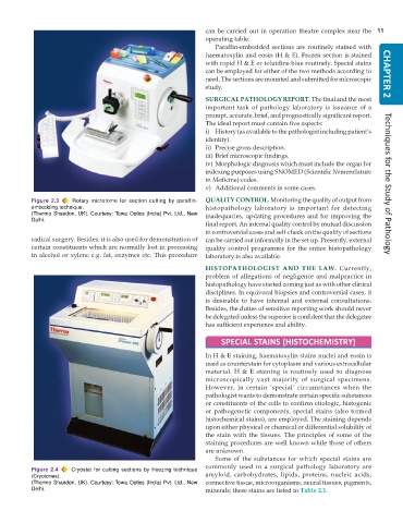
           Figure 2.3  Rotary microtome for section cutting by paraffin-  QUALITY CONTROL. Monitoring the quality of output from
           embedding technique.                                histopathology laboratory is important for detecting
           (Thermo Shandon, UK). Courtesy: Towa Optics (India) Pvt. Ltd., New  inadequacies, updating procedures and for improving the
           Delhi.
                                                               final report. An internal quality control by mutual discussion
                                                               in controversial cases and self-check on the quality of sections
           radical surgery. Besides, it is also used for demonstration of  can be carried out informally in the set up. Presently, external
           certain constituents which are normally lost in processing  quality control programme for the entire histopathology
           in alcohol or xylene e.g. fat, enzymes etc. This procedure  laboratory is also available.
                                                               HISTOPATHOLOGIST AND THE LAW.  Currently,
                                                               problem of allegations of negligence and malpractice in
                                                               histopathology have started coming just as with other clinical
                                                               disciplines. In equivocal biopsies and controversial cases, it
                                                               is desirable to have internal and external consultations.
                                                               Besides, the duties of sensitive reporting work should never
                                                               be delegated unless the superior is confident that the delegatee
                                                               has sufficient experience and ability.

                                                                    SPECIAL STAINS (HISTOCHEMISTRY)

                                                               In H & E staining, haematoxylin stains nuclei and eosin is
                                                               used as counterstain for cytoplasm and various extracellular
                                                               material. H & E staining is routinely used to diagnose
                                                               microscopically vast majority of surgical specimens.
                                                               However, in certain ‘special’ circumstances when the
                                                               pathologist wants to demonstrate certain specific substances
                                                               or constituents of the cells to confirm etiologic, histogenic
                                                               or pathogenetic components, special stains (also termed
                                                               histochemical stains), are employed. The staining depends
                                                               upon either physical or chemical or differential solubility of
                                                               the stain with the tissues. The principles of some of the
                                                               staining procedures are well known while those of others
                                                               are unknown.
                                                                  Some of the substances for which special stains are
                                                               commonly used in a surgical pathology laboratory are
           Figure 2.4  Cryostat for cutting sections by freezing technique
           (Cryotones).                                        amyloid, carbohydrates, lipids, proteins, nucleic acids,
           (Thermo Shandon, UK). Courtesy: Towa Optics (India) Pvt. Ltd., New  connective tissue, microorganisms, neural tissues, pigments,
           Delhi.                                              minerals; these stains are listed in Table 2.1.

