


Page 29 - Textbook of Pathology, 6th Edition
P. 29
13
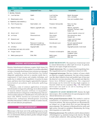
               TABLE 2.1: Contd...
               Stain                   Component/Tissue        Dyes                    Interpretation
           F. NEURAL TISSUES
           21. Luxol fast blue         Myelin                  Luxol fast blue,       Myelin: blue/green
                                                               cresyl violet          Cells: violet/pink              CHAPTER 2
           22. Bielschowsky’s silver   Axons                   Silver nitrate         Axon and neurofibrils: black
           G. PIGMENTS AND MINERALS
           23. Perl’s Prussian blue    Haemosiderin, iron      Potassium ferrocyanide  Ferric iron: blue
                                                                                      Nuclei: red
           24. Masson-Fontana          Melanin, argentaffin cells  Silver nitrate     Melanin, argentaffin,
                                                                                      chromaffin, lipofuscin: black
                                                                                      Nuclei: red
           25. Alizarin red S          Calcium                 Alizarin red S         Calcium deposits: orange red
           26. von Kossa               Mineralised bone        Silver nitrate,        Mineralised bone: black
                                                               safranin O             Osteoid: red
           27. Rubeanic acid           Copper                  Rubeanic acid          Copper: greenish-black
                                                                                      Nuclei: pale red
           28. Pigment extraction      Removal of formalin pig-  Alcoholic picric acid  Formalin pigment/malarial
                                       ment and malarial pigment                      pigment: removed                Techniques for the Study of Pathology
           29. Grimelius’              Argyrophil cells        Silver nitrate         Argyrophil granules: brown-black
           H. PROTEINS AND NUCLEIC ACIDS
           30. Feulgen reaction        DNA                     Potassium metabisulphite  DNA: red purple
                                                                                      Cytoplasm: green
           31. Methyl green-pyronin    DNA, RNA                Methyl green,          DNA: green-blue
                                                               pyronin-Y              RNA: red


                    ENZYME HISTOCHEMISTRY                      LIGHT MICROSCOPY. The usual type of microscope used
                                                               in clinical laboratories is called light microscope. In general,
           Enzyme histochemical techniques require fresh tissues for  there are two types of light microscopes:
           cryostat section and cannot be applied to paraffin-embedded  Simple microscope. This is a simple hand magnifying lens.
           sections or formalin-fixed tissues since enzymes are damaged  The magnification power of hand lens is from 2x to 200x.
           rapidly. Currently, enzyme histochemistry has limited  Compound microscope. This has a battery of lenses which
           diagnostic applications and not so popular, partly due to  are fitted in a complex instrument. One type of lens remains
           requirement of fresh tissues and complex technique, and  near the object (objective lens) and another type of lens near
           partly due to relative lack of specificity of reaction in many  the observer’s eye (eye piece lens). The eyepiece and objective
           cases, and hence have been largely superseded by immuno-  lenses have different magnification. The compound
           histochemical procedures and molecular pathology    microscope can be  monocular having single eyepiece or
           techniques.                                         binocular which has two eyepieces (Fig.  2.5). Multi-headed
              Presently, some of common applications of enzyme  microscopes  are used as an aid to teaching and for
           histochemistry in diagnostic pathology are in demonstration  demonstration purposes.
           of muscle related enzymes (ATPase) in myopathies,
           acetylcholinesterase in diagnosis of Hirschsprung’s disease,  VARIANTS OF LIGHT MICROSCOPY. Besides the light
           choloroacetate esterase for identification of myeloid cells and  microscopes, other modifications for special purposes in the
           mast cells, DOPA reaction for tyrosinase activity in  clinical laboratories are as under:
           melanocytes, endogenous dehydrogenase (requiring    Dark ground illumination (DGI). This method is used for
           nitroblue tetrazolium or NBT) for viability of cardiac muscle,  examination of unstained living microorganisms e.g.
           and acid and alkaline phosphatases.                 Treponema pallidum. The microorganisms are illuminated by
                                                               an oblique ray of light which does not pass through the
                                                               microorganism. The condenser is blackened in the centre and
                        BASIC MICROSCOPY
                                                               light passes through its periphery illuminating the living
                                                               microorganism on a glass slide.
           Microscope is the basic tool of the pathologist just as is the
           stethoscope for the physician and speculum for gynaecologist.  Polarising microscope. This method is used for demonstration
           It is an instrument which produces greatly enlarged images  of birefringence e.g. amyloid, foreign body, hair etc. The light
           of minute objects.                                  is made plane polarised. After passing through a disc, the

