


Page 368 - Textbook of Pathology, 6th Edition
P. 368
352   Laboratory Findings

            The diagnosis of IM is made by characteristic haemato-
            logic and serologic findings.
            1. HAEMATOLOGIC FINDINGS. Major abnormalities
            in blood are as under:
            i) TLC: There is a moderate rise in total white cell count
            (10,000-20,000/μl) during 2nd to 3rd week after infection.
            ii) DLC: There is an absolute lymphocytosis. The lympho-
            cytosis is due to rise in normal as well as atypical T
            lymphocytes. There is relative neutropenia.
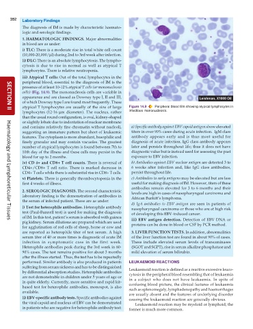
            iii) Atypical T cells: Out of the total lymphocytes in the
            peripheral blood, essential to the diagnosis of IM is the
            presence of at least 10-12% atypical T cells (or mononucleosis
            cells) (Fig. 14.9). The mononucleosis cells are variable in
            appearance and are classed as Downey type I, II and III,
            of which Downey type I are found most frequently. These
            atypical T lymphocytes are usually of the size of large  Figure 14.9  Peripheral blood film showing atypical lymphocytes in
            lymphocytes (12-16  μm diameter). The nucleus, rather  infectious mononucleosis.
     SECTION II
            than the usual round configuration, is oval, kidney-shaped
            or slightly lobate due to indentation of nuclear membrane
            and contains relatively fine chromatin without nucleoli,  a) Specific antibody against EBV capsid antigen show elevated
            suggesting an immature pattern but short of leukaemic  titers in over 90% cases during acute infection.  IgM class
            features. The cytoplasm is more abundant, basophilic and  antibody appears early and is thus most useful for
            finely granular and may contain vacuoles. The greatest  diagnosis of acute infection. IgG class antibody appears
            number of atypical lymphocytes is found between 7th to  later and persists throughout life; thus it does not have
            10th day of the illness and these cells may persist in the  diagnostic value but is instead used for assessing the past
            blood for up to 2 months.                            exposure to EBV infection.
            iv) CD 4+ and CD8+ T cell counts. There is reversal of  b) Antibodies against EBV nuclear antigen are detected 3 to
            CD4+/CD8+ T cell ratio. There is marked decrease in  6 weeks after infection and, like IgG class antibodies,
            CD4+ T cells while there is substantial rise in CD8+ T cells.  persist throughout life.
            v) Platelets. There is generally thrombocytopenia in the  c) Antibodies to early antigens may be elevated but are less
            first 4 weeks of illness.                            useful for making diagnosis of IM. However, titers of these
                                                                 antibodies remain elevated for 3 to 6 months and their
            2. SEROLOGIC DIAGNOSIS. The second characteristic    levels are high in cases of nasopharyngeal carcinoma and
            laboratory finding is the demonstration of antibodies in  African Burkitt’s lymphoma.
            the serum of infected patient. These are as under:
                                                                 d) IgA antibodies to EBV antigen are seen in patients of
            i) Test for heterophile antibodies. Heterophile antibody  nasopharyngeal carcinoma or those who are at high risk
            test (Paul-Bunnell test) is used for making the diagnosis  of developing this EBV-induced cancer.
            of IM. In this test, patient’s serum is absorbed with guinea  iii) EBV antigen detection. Detection of EBV DNA or
     Haematology and Lymphoreticular Tissues
            pig kidney. Serum dilutions are prepared which are used  proteins can be done in blood or CSF by PCR method.
            for agglutination of red cells of sheep, horse or cow and
            are reported as heterophile titer of test serum. A high  3. LIVER FUNCTION TESTS. In addition, abnormalities
            serum titer of 40 or more times is diagnostic of acute IM  of the liver function test are found in about 90% of cases.
            infection in symptomatic case in the first week.     These include elevated serum levels of transaminases
            Heterophile antibodies peak during the 3rd week in 80-  (SGOT and SGPT), rise in serum alkaline phosphatase and
            90% cases. The test remains positive for about 3 months  mild elevation of serum bilirubin.
            after the illness started.  Thus, the test has to be repeatedly
            performed. Similar antibody is also produced in patients  LEUKAEMOID REACTIONS
            suffering from serum sickness and has to be distinguished  Leukaemoid reaction is defined as a reactive excessive leuco-
            by differential absorption studies. Heterophile antibodies  cytosis in the peripheral blood resembling that of leukaemia
            are not demonstrable in children under 5 years of age or  in a subject who does not have leukaemia. In spite of
            in quite elderly. Currently, more sensitive and rapid kit-  confusing blood picture, the clinical features of leukaemia
            based test for heterophile antibodies, monospot, is also  such as splenomegaly, lymphadenopathy and haemorrhages
            available.                                         are usually absent and the features of underlying disorder
            ii) EBV-specific antibody tests. Specific antibodies against  causing the leukaemoid reaction are generally obvious.
            the viral capsid and nucleus of EBV can be demonstrated  Leukaemoid reaction may be myeloid or lymphoid; the
            in patients who are negative for heterophile antibody test:  former is much more common.

