


Page 364 - Textbook of Pathology, 6th Edition
P. 364
348 4. Acute haemorrhage, internal or external.
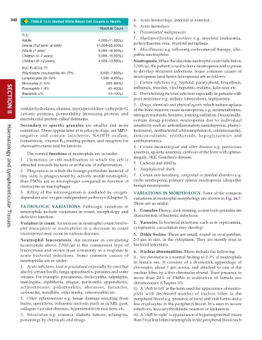
TABLE 14.2: Normal White Blood Cell Counts in Health.
5. Acute haemolysis.
Absolute Count
6. Disseminated malignancies.
TLC 7. Myeloproliferative disorders e.g. myeloid leukaemia,
Adults 4,000–11,000/μl polycythaemia vera, myeloid metaplasia.
Infants (Full term, at birth) 10,000–25,000/μl
Infants (1 year) 6,000–16,000/μl 8. Miscellaneous e.g. following corticosteroid therapy, idio-
Children (4–7 years) 5,000–15,000/μl pathic neutrophilia.
Children (8–12 years) 4,500–13,500/μl Neutropenia. When the absolute neutrophil count falls below
2,500/μl, the patient is said to have neutropenia and is prone
DLC IN ADULTS
Polymorphs (neutrophils) 40–75% 2,000–7,500/μl to develop recurrent infections. Some common causes of
Lymphocytes 20–50% 1,500–4,000/μl neutropenia (and hence leucopenia) are as follows:
Monocytes 2–10% 200–800/μl 1. Certain infections e.g. typhoid, paratyphoid, brucellosis,
Eosinophils 1–6% 40–400/μl influenza, measles, viral hepatitis, malaria, kala-azar etc.
Basophils <l% 10–100/μl 2. Overwhelming bacterial infections especially in patients with
poor resistance e.g. miliary tuberculosis, septicaemia.
3. Drugs, chemicals and physical agents which induce aplasia
contain hydrolases, elastase, myeloperoxidase, cathepsin-G, of the bone marrow cause neutropenia, e.g. antimetabolites,
cationic proteins, permeability increasing protein, and nitrogen mustards, benzene, ionising radiation. Occasionally,
SECTION II
microbicidal protein called defensins. certain drugs produce neutropenia due to individual
Secondary or specific granules are smaller and more sensitivity such as: anti-inflammatory (amidopyrine, phenyl-
numerous. These appear later at myelocyte stage, are MPO- butazone), antibacterial (chloramphenicol, cotrimoxazole),
negative and contain lactoferrin, NADPH oxidase, anticonvulsants, antithyroids, hypoglycaemics and
histaminase, vitamin B binding protein, and receptors for antihistaminics.
12
chemoattractants and for laminin. 4. Certain haematological and other diseases e.g. pernicious
The normal functions of neutrophils are as under: anaemia, aplastic anaemia, cirrhosis of the liver with spleno-
1. Chemotaxis or cell mobilisation in which the cell is megaly, SLE, Gaucher’s disease.
attracted towards bacteria or at the site of inflammation. 5. Cachexia and debility.
2. Phagocytosis in which the foreign particulate material of 6. Anaphylactoid shock.
tiny sizes is phagocytosed by actively motile neutrophils; 7. Certain rare hereditary, congenital or familial disorders e.g.
thus PMNs act as microphages compared to function of cyclic neutropenia, primary splenic neutropenia, idiopathic
monocytes as macrophages. benign neutropenia.
3. Killing of the microorganism is mediated by oxygen- VARIATIONS IN MORPHOLOGY. Some of the common
dependent and oxygen-independent pathways (Chapter 3). variations in neutrophil morphology are shown in Fig. 14.7.
These are as under:
PATHOLOGIC VARIATIONS. Pathologic variations in
neutrophils include variations in count, morphology and 1. Granules. Heavy, dark staining, coarse toxic granules are
defective function. characteristic of bacterial infections.
Variation in count. An increase in neutrophil count (neutro- 2. Vacuoles. In bacterial infections such as in septicaemia,
phil leucocytosis or neutrophilia) or a decrease in count cytoplasmic vacuolation may develop.
Haematology and Lymphoreticular Tissues
(neutropenia) may occur in various diseases. 3. Döhle bodies. These are small, round or oval patches,
Neutrophil leucocytosis. An increase in circulating 2-3 μm in size, in the cytoplasm. They are mostly seen in
neutrophils above 7,500/μl is the commonest type of bacterial infections.
leucocytosis and occurs most commonly as a response to 4. Nuclear abnormalities. These include the following:
acute bacterial infections. Some common causes of i) Sex chromatin is a normal finding in 2-3% of neutrophils
neutrophilia are as under: in female sex. It consists of a drumstick appendage of
1. Acute infections, local or generalised, especially by cocci but chromatin, about 1 μm across, and attached to one of the
also by certain bacilli, fungi, spirochaetes, parasites and some nuclear lobes by a thin chromatin strand. Their presence in
viruses. For example: pneumonia, cholecystitis, salpingitis, more than 20% of PMNs is indicative of female sex
meningitis, diphtheria, plague, peritonitis, appendicitis, chromosomes (Chapter 10).
actinomycosis, poliomyelitis, abscesses, furuncles, ii) A ‘shift-to-left’ is the term used for appearance of neutro-
carbuncles, tonsillitis, otitis media, osteomyelitis etc. phils with decreased number of nuclear lobes in the
2. Other inflammations e.g. tissue damage resulting from peripheral blood e.g. presence of band and stab forms and a
burns, operations, ischaemic necrosis (such as in MI), gout, few myelocytes in the peripheral blood. It is seen in severe
collagen-vascular diseases, hypersensitivity reactions etc. infections, leucoerythroblastic reaction or leukaemia.
3. Intoxication e.g. uraemia, diabetic ketosis, eclampsia, iii) A ‘shift-to-right’ is appearance of hypersegmented (more
poisonings by chemicals and drugs. than 5 nuclear lobes) neutrophils in the peripheral blood such

