


Page 365 - Textbook of Pathology, 6th Edition
P. 365
349


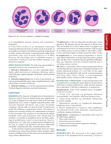
           Figure 14.7  Common variations in neutrophil morphology.


           as in megaloblastic anaemia, uraemia, and sometimes in  T lymphocytes i.e. thymus-dependent lymphocytes, which  CHAPTER 14
           leukaemia.                                          mature in the thymus and are also known as thymocytes.
           iv) Pelger-Huët anomaly is an uncommon autosomal    They are mainly involved in direct action on antigens and
           dominant inherited disorder in which nuclei in majority of  are therefore involved in cell-mediated immune (CMI) reaction
           neutrophils are distinctively bilobed (spectacle-shaped) and  by its subsets such as cytotoxic (killer) T cells (CD3+), CD8+
           coarsely staining chromatin. Acquired pseudo-Pelger-Huët  T cells, and delayed hypersensitivity reaction by CD4+ T cells.
           abnormality may occur in acute infections or in MDS.  B lymphocytes i.e. bone marrow-dependent or bursa-
           However, the physiologic role of multilobed nucleus of  equivalent lymphocytes  as well as their derivatives, plasma
           neutrophils is unknown and the bilobed anomaly is an  cells, are the source of specific immunoglobulin antibodies.
           innocuous condition.                                They are, therefore, involved in humoral immunity (HI) or
                                                               circulating immune reactions.
           DEFECTIVE FUNCTIONS. The following abnormalities in
           neutrophil function may sometimes be found:         NK cells i.e. natural killer cells are those lymphocytes which
                                                               morphologically have appearance of lymphocytes but do not
           1. Defective chemotaxis e.g. in a rare congenital abnor-  possess functional features of T or B cells. As the name
           mality called lazy-leucocyte syndrome; following cortico-  indicates they are identified with ‘natural’ or innate immunity
           steroid therapy, aspirin ingestion, alcoholism, and in myeloid  and bring about direct ‘killing’ of microorganisms
           leukaemia.                                          (particularly certain viruses) or lysis of foreign body.
           2. Defective phagocytosis due to lack of opsonisation e.g.                                                 Disorders of Leucocytes and Lymphoreticular Tissues
           in hypogammaglobulinaemia, hypocomplementaemia, after  PATHOLOGIC VARIATIONS. A rise in the absolute count
           splenectomy, in sickle cell disease.                of lymphocytes exceeding the upper limit of normal (above
           3. Defective killing e.g. in chronic granulomatous disease,  4,000/μm) is termed lymphocytosis, while absolute lympho-
           Chédiak-Higashi syndrome, myeloid leukaemias.       cyte count below 1,500/μm is referred to as lymphopenia.
                                                               Lymphocytosis. Some of the common causes of lympho-
                                                               cytosis are as under:
           Lymphocytes
                                                               1. Certain acute infections e.g. pertussis, infectious
           MORPHOLOGY. Majority of lymphocytes in the peripheral  mononucleosis, viral hepatitis, infectious lymphocytosis.
           blood are small (9-12 μm in diameter) but large lymphocytes  2. Certain chronic infections e.g. brucellosis, tuberculosis,
           (12-16 μm in diameter) are also found. Both small and large  secondary syphilis.
           lymphocytes have round or slightly indented nucleus with  3. Haematopoietic disorders e.g. lymphocytic leukaemias,
           coarsely-clumped chromatin and scanty basophilic    lymphoma, heavy chain disease.
           cytoplasm. Plasma cells are derived from B lymphocytes  4. Relative lymphocytosis is found in viral exanthemas,
           under the influence of appropriate stimuli. The nucleus of  convalescence from acute infections, thyrotoxicosis,
           plasma cell is eccentric and has cart-wheel pattern of clumped  conditions causing neutropenia.
           nuclear chromatin. The cytoplasm is characteristically deeply  Lymphopenia. Lymphopenia is uncommon and occurs in
           basophilic with a pale perinuclear zone. Plasma cells are  the following conditions:
           normally not present in peripheral blood but their patho-  1. Most acute infections.
           logical proliferation occurs in myelomatosis. Reactive  2. Severe bone marrow failure.
           lymphocytes (or Turk cells or plasmacytoid lymphocytes)  3. Corticosteroid and immunosuppressive therapy.
           are seen in certain viral infections and have sufficiently  4. Widespread irradiation.
           basophilic cytoplasm that they resemble plasma cells.
               As discussed in Chapter 4, functionally, there are 3 types
           of lymphocytes and possess distinct surface markers called  Monocytes
           clusters of differentiation (CD) which aid in identification of  MORPHOLOGY.  The monocyte is the largest mature
           stage of their differentiation:                     leucocyte in the peripheral blood measuring 12-20 μm in

