


Page 382 - Textbook of Pathology, 6th Edition
P. 382
366
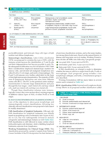
TABLE 14.6: FAB Classification of ALL.
A. MORPHOLOGIC CRITERIA
Fab Class Percent Cases Morphology Cytochemistry
L1: Childhood-ALL More common Homogeneous small lymphoblasts; scanty PAS ±
(B-ALL, and T-ALL) in children cytoplasm, regular round nuclei, Acid phosphatase ±
inconspicuous nucleoli
L2: Adult-ALL More frequent Heterogeneous lymphoblasts; variable amount PAS ±
(mostly T-ALL) in adults of cytoplasm, irregular or cleft nuclei, large nucleoli Acid phosphatase ±
L3: Burkitt type-ALL Uncommon Large homogeneous lymphoblasts; round nuclei, PAS –
(B-ALL) prominent nucleoli, cytoplasmic vacuolation Acid phosphatase –
B. CYTOGENETIC AND IMMUNOLOGIC CRITERIA
Subtype Incidence Markers FAB Subtype Cytogenetic Abnormalities
Pre-B ALL 75% CD10+ (90%), TdT+ L1, L2 t(9;22) i.e. Philadelphia+ALL
B cell ALL 5% CD10+ (50%), TdT- L3 t(8;14) (Burkitt’s leukaemia)
T cell ALL 20% CD10+ (30%), TdT+ L1, L2 14q11
(TdT= terminal deoxynucleotidyl transferase)
SECTION II
poorly-differentiated, and histiocytic (large cells) types of both all previous classification systems, and as the name implies,
nodular and diffuse lymphomas. has strong clinical relevance. Based on the natural history of
disease and long-term survival studies, Working Formula-
Immunologic classifications. Lukes-Collins classification
(1974) was proposed to correlate the type of NHL with the tions divides all NHLs into following 3 prognostic groups:
immune system because the identification of T and B-cells Low-grade NHL: 5-year survival 50-70%
and their subpopulations had become possible in early 70s. Intermediate-grade NHL: 5-year survival 35-45%
Its subsequent modification was Kiel classification (1981). Both High-grade NHL: 5-year survival 25-35%.
these classifications employed immunologic markers for In this classification, no attempt is made to determine
tumour cells, and divided all malignant lymphomas into whether the tumour cells have origin from B-cells, T-cells or
either B-cell or T-cell origin, and rarely of macrophages. The macrophages. Each prognostic group includes a few
B and T-cell tumours were further subdivided on the basis morphologic subtypes, and lastly, a miscellaneous group is
of their light microscopic characteristics. The majority of NHL also described.
were B lymphocyte derivatives and arise from follicular Working Formulations still has many takers in several
centre cells (FCC). The FCC in the germinal centre undergo centres and is retained in Table 14.7.
transformation to become large immunoblasts and pass
through the four stages—small cleaved cells and large cleaved REAL classification (1994). International Lymphoma Study
cells, small non-cleaved cells and large non-cleaved cells. Group (Harris et al) proposed another classification called
Though these classification schemes were immuno-
logically correct, they were unclear about varying prognosis TABLE 14.7: Classiifcation of NHL-Working Formulations
of different clinical types of NHL of either B-cell or T-cell for Clinical Usage (1982).
Haematology and Lymphoreticular Tissues
origin.
I. LOW-GRADE
II. OLD CLINICOPATHOLOGIC CLASSIFICATIONS. In A) Small lymphocytic
view of the objections to above pure morphologic and B) Follicular, predominantly small cleaved cell
immunologically correct classifications, following two C) Follicular, mixed small and large cleaved cell
clinically relevant classifications were proposed which cannot II. INTERMEDIATE-GRADE
be readily abandoned: D) Follicular, predominantly large cell
FAB classification of lymphoid leukaemia. Although old, E) Diffuse, small cleaved cell
FAB classification for lymphoid leukaemia was initially F) Diffuse, mixed small and large cell
based on morphology and cytochemistry into 3 types of ALL G) Diffuse, large, cell
(L1 to L3), but was subsequently revised to include III. HIGH-GRADE
cytogenetic and immunologic features as well (Table 14.6). H) Large cell, immunoblastic
FAB classification is still followed in many centres where I) Lymphoblastic
both pathologists and clinicians stick to labelling lymphoid J) Small non-cleaved cell (Burkitt’s)
leukaemia separate from lymphomas. IV. MISCELLANEOUS
Working Formulations for Clinical Usage (1982). This 1. Adult T-cell leukaemia/lymphoma
classification proposed by a panel of experts from National 2. Cutaneous T-cell lymphoma
Cancer Institute of the US incorporates the best features of 3. Histiocytic (Histiocytic medullary reticulosis)

