


Page 386 - Textbook of Pathology, 6th Edition
P. 386
370


     SECTION II


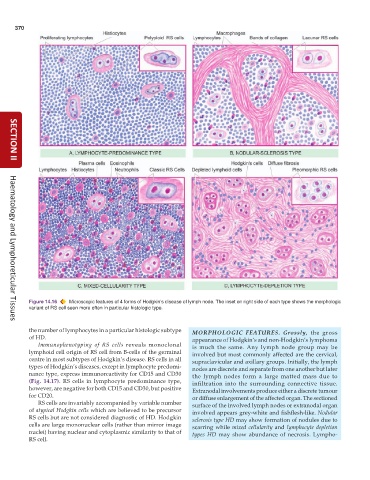
           Figure 14.16  Microscopic features of 4 forms of Hodgkin’s disease of lymph node. The inset on right side of each type shows the morphologic
           variant of RS cell seen more often in particular histologic type.

     Haematology and Lymphoreticular Tissues
           the number of lymphocytes in a particular histologic subtype  MORPHOLOGIC FEATURES.  Grossly, the gross
           of HD.                                                appearance of Hodgkin’s and non-Hodgkin’s lymphoma
              Immunophenotyping of RS cells reveals monoclonal   is much the same. Any lymph node group may be
           lymphoid cell origin of RS cell from B-cells of the germinal  involved but most commonly affected are the cervical,
           centre in most subtypes of Hodgkin’s disease. RS cells in all  supraclavicular and axillary groups. Initially, the lymph
           types of Hodgkin’s diseases, except in lymphocyte predomi-  nodes are discrete and separate from one another but later
           nance type, express immunoreactivity for CD15 and CD30  the lymph nodes form a large matted mass due to
           (Fig. 14.17). RS cells in lymphocyte predominance type,  infiltration into the surrounding connective tissue.
           however, are negative for both CD15 and CD30, but positive  Extranodal involvements produce either a discrete tumour
           for CD20.                                             or diffuse enlargement of the affected organ. The sectioned
              RS cells are invariably accompanied by variable number  surface of the involved lymph nodes or extranodal organ
           of atypical Hodgkin cells which are believed to be precursor  involved appears grey-white and fishflesh-like. Nodular
           RS cells but are not considered diagnostic of HD. Hodgkin  sclerosis type HD may show formation of nodules due to
           cells are large mononuclear cells (rather than mirror image  scarring while mixed cellularity and  lymphocyte depletion
           nuclei) having nuclear and cytoplasmic similarity to that of  types  HD may show abundance of necrosis. Lympho-
           RS cell.

