


Page 385 - Textbook of Pathology, 6th Edition
P. 385
6. Immune abnormalities: Since lymphoid neoplasms arise The salient features of the 4 histologic subtypes of HD 369
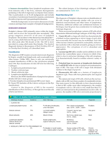
from immune cells of the body, immune derangements are summarised in Table 14.9.
pertaining to the cell of origin may accompany these cancers.
This is particularly so in B-cell malignancies and include Reed-Sternberg Cell
occurrence of autoimmune haemolytic anaemia, autoimmune
thrombocytopenia and hypogammaglobulinaemia. The diagnosis of Hodgkin’s disease rests on identification of
RS cells, though uncommonly similar cells can occur in
With this background, we now turn to discussion of some infectious mononucleosis and other forms of lymphomas.
common and important examples of lymphoid malignancies.
Therefore, additional cellular and architectural features of
the biopsy must be given due consideration for making the
HODGKIN’S DISEASE
histologic diagnosis.
Hodgkin’s disease (HD) primarily arises within the lymph There are several morphologic variants of RS cells which
nodes and involves the extranodal sites secondarily. This characterise different histologic subtypes of HD (Fig. 14.16):
group comprises about 8% of all cases of lymphoid 1. Classic RS cell is a large cell which has characteristically
neoplasms. The incidence of the disease has bimodal peaks— a bilobed nucleus appearing as mirror image of each other
one in young adults between the age of 15 and 35 years and but occasionally the nucleus may be multilobed. Each lobe
the other peak after 5th decade of life. The HD is more of the nucleus contains a prominent, eosinophilic, inclusion- CHAPTER 14
prevalent in young adult males than females. The classical like nucleolus with a clear halo around it, giving an owl-eye
diagnostic feature is the presence of Reed-Sternberg (RS) cell appearance. The cytoplasm of cell is abundant and
(or Dorothy-Reed-Sternberg cell) (described later).
amphophilic.
Classification 2. Lacunar type RS cell is smaller and in addition to above
features has a pericellular space or lacuna in which it lies,
The diagnosis of HD requires accurate microscopic diagnosis
by biopsy, usually from lymph node, and occasionally from which is due to artefactual shrinkage of the cell cytoplasm.
other tissues. Unlike NHL, there is only one universally It is characteristically found in nodular sclerosis variety of
accepted classification of HD i.e. Rye classification adopted HD.
since 1966. Rye classification divides HD into the following 3. Polyploid type (or popcorn or lymphocytic-histiocytic
4 subtypes: i.e. L and H) RS cells are seen in lymphocyte predominance
1. Lymphocyte-predominance type type of HD. This type of RS cell is larger with lobulated
2. Nodular-sclerosis type nucleus in the shape of popcorn.
3. Mixed-cellularity type 4. Pleomorphic RS cells are a feature of lymphocyte
4. Lymphocyte-depletion type. depletion type. These cells have pleomorphic and atypical
However, the WHO classification of lymphoid neoplasms nuclei.
divides HD into 2 main groups: The nature and origin of RS cells, which are the real neo- Disorders of Leucocytes and Lymphoreticular Tissues
I. Nodular lymphocyte-predominant HD (a new type). plastic cells in HD, have been a matter of considerable debate.
II. Classic HD (includes all the 4 above subtypes in the Rye One main reason for this difficulty in their characterisation
classification). is that in HD, unlike most other malignancies, the number
Central to the diagnosis of HD is the essential of neoplastic cells (i.e. RS cells) is very small (less than 5%)
identification of Reed-Sternberg cell though this is not the sole which are interspersed in the predominant reactive cells. In
criteria (see below). general, the number of RS cells is inversely proportional to
TABLE 14.9: Modified WHO Classification of Hodgkin’s Disease.
Histologic Subtype Incidence Main Pathology RS Cells Prognosis
I. CLASSIC HD
Lymphocyte-predominance 5% Proliferating lymphocytes, Few, classic and polyploid Excellent
a few histiocytes type, CD15–, CD30–, CD20+
Nodular sclerosis 70% Lymphoid nodules, Frequent, lacunar type, Very good
collagen bands CD15+, CD30+
Mixed cellularity 22% Mixed infiltrate Numerous, classic type, Good
CD15+, CD30+
Lymphocyte-depletion 1% Scanty lymphocytes, Numerous, Poor
(Diffuse fibrotic and atypical histiocytes, fibrosis pleomorphic type,
reticular variants) CD15+, CD30+
II. NODULAR LYMPHOCYTE-PREDOMINANT HD
2% Proliferation of small lympho- Sparse number of RS cells, Chronic
cytes, nodular pattern of growth CD45+, EMA+, CD15-, CD30- relapsing, may
transform into
large B cell NHL

