


Page 384 - Textbook of Pathology, 6th Edition
P. 384
368
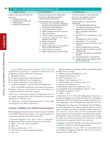
TABLE 14.8: WHO Classification of Lymphoid Malignancies (1999).
Hodgkin’s Disease B Cell Malignancies T Cell Malignancies
Nodular lymphocytic predominant HD Precursor (Immature) B-cell malignancies Precursor (Immature) T-cell malignancies
Classic HD Precursor B lymphoblastic leukaemia/ Precursor T lymphoblastic lymphoma/
1. Nodular sclerosis HD lymphoma (precursor B-cell ALL) leukaemia (Precursor T-cell ALL)
2. Lymphocytic rich classic HD Peripheral (Mature) B-cell malignancies Peripheral (Mature) T-cell and NK-cell
3. Mixed cellularity 1. B-cell CLL/SLL (chronic lymphocytic malignancies
4. Lymphocytic depletion HD
leukaemia/small lymphocytic lymphoma) 1. T-cell prolymphocytic leukaemia
2. B-cell prolymphocytic leukaemia 2. T-cell granular lymphocytic leukaemia
3. Lymphoplasmacytic lymphoma 3. Aggressive NK cell leukaemia
4. Splenic marginal zone B-cell lymphoma 4. Adult T-cell lymphoma/leukaemia
5. Hairy cell leukaemia (HTLV-I +)
6. Plasma cell myeloma/plasmacytoma 5. Extranodal NK/T-cell lymphoma, nasal
7. Extranodal marginal zone B-cell type
lymphoma, MALT type 6. Enteropathy-type T-cell lymphoma
8. Mantle cell lymphoma 7. Hepatosplenic T-cell lymphoma
9. Follicular lymphoma 8. Subcutaneous panniculitis like T-cell
lymphoma
10. Nodal marginal zone B-cell lymphoma
(Monocytoid B-cell lymphoma) 9. Mycosis fungoides/Sézary syndrome
SECTION II
11. Diffuse large B-cell lymphoma 10. Anaplastic large T lymphoma, primary
12. Burkitt’s lymphoma/Burkitt cell leukaemia cutaneous type
11. Peripheral T-cell lymphoma, not otherwise
specified
12. Angioimmunoblastic T-cell lymphoma
13. Anaplastic large T-cell lymphoma (ALCL),
primary systemic type
As per WHO classification scheme (Table 14.8), all Relative frequency of subtypes within various NHLs listed
lymphoid neoplasms (i.e. lymphoid leukaemias and in Table 14.8 is as under:
lymphomas) fall into following 5 categories: i) Diffuse large B cell lymphoma = 31%
I. Hodgkin’s disease ii) Follicular lymphoma = 22%
II. Precursor (Immature) B-cell malignancies iii) MALT lymphoma = 8%
III. Peripheral (Mature) B-cell malignancies iv) Mature T cell lymphoma = 8%
IV. Precursor (Immature) T-cell malignancies v) Small lymphocytic lymphoma (SLL) = 7%
V. Peripheral (Mature) T-cell and NK–cell malignancies vi) Mantle cell lymphoma = 6%
Thus, in the WHO classification of lymphoid neoplasms, vii)Mediastinal large B cell lymphoma = 2.5%
Hodgkin’s disease stands distinctive; remaining four viii) Anaplastic large cell lymphoma (ALCL) = 2.5%
categories listed above fall into 2 groups: ix) Burkitt’s lymphoma = 2.5%
x) Others = ~10%
1) Precursor or immature lymphoid malignancies of B or T
cell origin, meaning blastic type of leukaemias-lymphomas 2. Incidence of B, T, NK cell malignancies: Majority of
Haematology and Lymphoreticular Tissues
(mainly B or T-cell ALL); and lymphoid malignancies are of B cell origin (75% of lymphoid
leukaemias and 90% of lymphomas) while remaining are T
2) Peripheral or mature malignancies of B or T cell origin cell malignancies; NK-cell lymphomas-leukaemias are rare.
(meaning CLL and other lymphomas).
3. Diagnosis: The diagnosis of lymphoma (both Hodgkin’s
and non-Hodgkin’s) can only be reliably made on exami-
GENERAL COMMENTS ON LYMPHOID MALIGNANCIES
nation of lymph node biopsy. While the initial diagnosis of
Before plunging into discussion of various common examples ALL and CLL can be made on CBC examination, bone
in the WHO classification system, a few general aspects on marrow biopsy is done for genetic and immunologic studies.
lymphoid neoplasms need to be understood: Subsequently, clinical chemistry, electrophoresis and tests
for organ involvement including CSF examination if CNS
1. Overall frequency: Five major forms of lymphoid involvement is suspected, need to be carried out.
malignancies and their relative frequency are as under:
i) NHL= 62%, most common lymphoma 4. Staging: In both HD and NHL, Ann Arbor staging is done
ii) HD= 8% for proper evaluation and planning treatment.
iii) Plasma cell disorders = 15% 5. Ancillary studies: CT scan, PET scan and gallium scan
iv) CLL= 9%, most common lymphoid leukaemia are additional imaging modalities which can be used in
v) ALL= 4% staging HD and NHL cases.

