


Page 387 - Textbook of Pathology, 6th Edition
P. 387
ii) Lacunar type RS cells: Characteristic lacunar type of RS  371
                                                                 cells with distinctive pericellular halo are present. These
                                                                 cells appear lacunar due to the shrinkage of cytoplasm in
                                                                 formalin-fixed tissue. The pericellular halo is not seen if
                                                                 the tissue is fixed in Zenker’s fluid.
                                                                    In addition to these 2 characteristics, the nodules
                                                                 between the fibrous septa consist predominantly of
                                                                 lymphocytes and macrophages, sometimes with foci of
                                                                 necrosis.
                                                                 3. Mixed-cellularity type (Fig. 14.16,C). This form of HD
                                                                 generally replaces the entire affected lymph nodes by
                                                                 heterogeneous mixture of various types of apparently
                                                                 normal cells. These include proliferating lymphocytes,
                                                                 histiocytes, eosinophils, neutrophils and plasma cells.
                                                                 Some amount of fibrosis and focal areas of necrosis are
                                                                 generally present. Typical RS cells are frequent (Fig. 14.18,  CHAPTER 14
                                                                 B).
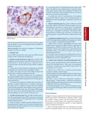
           Figure 14.17  RS cells showing positive immunostaining for  CD15,
           a B-cell marker.                                      4. Lymphocyte-depletion type (Fig. 14.16,D). In this type
                                                                 of HD, the lymph node is depleted of lymphocytes. There
                                                                 are two variants of lymphocyte-depletion HD:
            matous involvement of the liver, spleen and other organs
            may be diffuse or may form spherical masses similar to  i) Diffuse fibrotic variant is hypocellular and the entire
            metastatic carcinoma.                                lymph node is replaced by diffuse fibrosis, appearing as
                                                                 homogeneous, fibrillar hyaline material. The area of
            Microscopically, the criteria for diagnosis of histologic  hyalinosis contains some lymphocytes, atypical histiocytes
            subtypes of HD are as under:                         (Hodgkin cells), and numerous typical and atypical
            I.  CLASSIC HD                                       (pleomorphic) RS cells.
                                                                 ii) Reticular variant is much more cellular and consists of
            As per WHO classification, classic group of HD includes
            4 types of HD of older Rye classification:           large number of atypical pleomorphic histiocytes, scanty
                                                                 lymphocytes and a few typical RS cells.
            1. Lymphocyte-predominance type (Fig. 14.16,A). The
            lymphocyte-predominance type of HD is characterised by  II. NODULAR LYMPHOCYTE-PREDOMINANT HD
            proliferation of small lymphocytes admixed with a    This is a newly described entity which is distinct from the
            varying number of histiocytes forming nodular or diffuse  classic HD described above. This type was previously  Disorders of Leucocytes and Lymphoreticular Tissues
            pattern.                                             included in lymphocyte predominant type of HD. Its
            i) Nodular form is characterised by replacement of nodal  peculiarities are as under:
            architecture by numerous large neoplastic nodules.   i)  These cases of HD have a nodular growth pattern
            ii) Diffuse form does not have discernible nodules but  (similar to nodular sclerosis type).
            instead there is diffuse proliferation of cells.     ii) Like lymphocyte-predominant pattern of classic type,
               However, currently nodular form of lymphocyte     there is predominance of small lymphocytes with  sparse
            predominent HD has been categorised separately due to  number of RS cells.
            its distinct immunophenotyping features and prognosis  iii) These cases of HD have distinctive immuno-
            (discussed below).                                   phenotyping—CD45 positive, epithelial membrane
               For making the diagnosis, definite demonstration of  antigen (EMA) positive but negative for the usual markers
            RS cells is essential which are few in number, requiring a  for RS cells (CD15 and CD30 negative).
            thorough search. In addition to typical RS cells, polyploid  iv) Though generally it has a chronic relapsing course,
            variant having polyploid, and twisted nucleus (popcorn-  but some cases of this type of HD may transform into large
            like) may be found in some cases. This type of HD usually  B-cell NHL.
            does not show other cells like plasma cells, eosinophils
            and neutrophils, nor are necrosis or fibrosis seen.  Clinical Features

            2. Nodular-sclerosis type (Fig. 14.16,B). Nodular sclero-  Hodgkin’s disease is particularly frequent among young and
            sis is the most frequent type of HD, seen more commonly  middle-aged adults. All histologic subtypes of HD, except
            in women than in men. It is characterised by two essential  the nodular sclerosis variety, are more common in males.
            features (Fig. 14.18, A):                          The disease usually begins with superficial lymph node
            i) Bands of collagen: Variable amount of fibrous tissue is  enlargement and subsequently spreads to other lymphoid
            characteristically present in the involved lymph nodes.  and non-lymphoid structures.
            Occasionally, the entire lymph node may be replaced by  1. Most commonly, patients present with painless, movable
            dense hyalinised collagen.
                                                               and firm  lymphadenopathy. The cervical and mediastinal

