


Page 401 - Textbook of Pathology, 6th Edition
P. 401
diagnosed in asymptomatic healthy aging population—1%                                                    385
           at 50 years of age and in 10% individuals older than 75 years.
           This makes it the most common form of plasma cell dyscrasia.
           The defining criteria for MGUS are as under:
           i) M-protein in serum <3 gm/dl
           ii) Marrow plasmacytosis <10%
           iii) No evidence of other B-cell proliferative disorder
           iv) Absence of myeloma-related end-organ tissue damage
           (i.e. absence of lytic bone lesions, high calcium level,
           anaemia).
              The condition needs to be cautiously distinguished from
           myeloma and long term follow-up is required. In fact, MGUS
           has been considered as a forerunner of multiple myeloma in
           many studies because MGUS has been found to have the
           same chromosomal abnormalities as in myeloma.
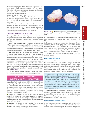
                                                               Figure 14.28  Metastatic carcinomatous deposits in the matted mass
           LYMPH NODE METASTATIC TUMOURS                       of lymph nodes. There are areas of necrosis in the circumscribed nodular  CHAPTER 14
                                                               areas.
           The regional lymph nodes draining the site of a primary
           malignant tumour are commonly enlarged. This enlargement  i) Demonstration of common antigens on these cells by
           may be due to benign reactive hyperplasia or metastatic tumour  immunohistochemical stains for S-100 protein, CD1a and HLA-
           deposits.                                           DR.
           1. Benign reactive hyperplasia, as already discussed (page  ii) Electron microscopic demonstration of histiocytosis-X bodies
           343), is due to immunologic reaction by the lymph node in  or Birbeck granules in the cytoplasm. These are rod-shaped
           response to tumour-associated antigens. It may be expressed  structures having dilated tennis-racket like terminal end.
           as sinus histiocytosis, follicular hyperplasia, plasmacytosis  Their function is not known but they arise from receptor-
           and occasionally may show non-caseating granulomas.  mediated endocytosis of langerin found in human epidermal
                                                               cells, a protein involved in Birbeck granule biosynthesis.
           2. Metastatic deposits in regional lymph nodes occur most  The three disorders included in the group are briefly
           commonly from carcinomas and malignant melanoma.    considered below.
           Sarcomas often disseminate via haematogenous route but
           uncommonly may metastasise to the regional lymph nodes.  Eosinophilic Granuloma
           Metastatic tumour cells from the primary malignant tumour
           are drained via lymphatics into the subcapsular sinuses  Unifocal eosinophilic granuloma is more common (60%) than
           initially but subsequently the lymph node stroma is also  the multifocal variety which is often a component of Hand-  Disorders of Leucocytes and Lymphoreticular Tissues
           invaded. The pushing margins of advancing metastatic  Schüller-Christian disease (described below). Most of the
           tumour in stroma of lymph node is characteristically well  patients are children and young adults, predominantly males.
           demarcated. Areas of necrosis are frequent in metastatic carci-  The condition commonly presents as a solitary osteolytic
           nomas (Fig. 14.28).                                 lesion in the femur, skull, vertebrae, ribs and pelvis. The
              The morphologic features of primary malignant tumour  diagnosis requires biopsy of the lytic bone lesion.
           are recapitulated in metastatic tumour in lymph nodes.
                                                                 Microscopically, the lesion consists largely of closely-
                                                                 packed aggregates of macrophages admixed with variable
                     HISTIOCYTIC NEOPLASMS:
                                                                 number of eosinophils (Fig. 14.29). The macrophages
               LANGERHANS’ CELL HISTIOCYTOSIS                    contain droplets of fat or a few granules of brown pigment
                                                                 indicative of phagocytic activity. A few multinucleate
           Langerhans’ cell histiocytosis (LCH) is a group of rare  macrophages may also be seen. The cytoplasm of these
           malignant proliferations of dendritic cells or macrophages  macrophages may contain rod-shaped inclusions called
           and includes three clinicopathologically related conditions  histiocytosis-X bodies or Birbeck granules, best seen by
           ocurring in children: eosinophilic granuloma, Hand-Schüller-  electron microscopy.
           Christian disease and Letterer-Siwe syndrome. Earlier, this
           group was referred to as histiocytosis-X but now following  Clinically, unifocal eosinophilic granuloma is a benign
           facts about this group are known:                   disorder. The bony lesion remains asymptomatic until the
              Firstly, histiocytosis-X are not proliferations of unknown  erosion of the bone causes pain or fracture. Spontaneous
           origin (X-for unknown) but proliferating cells are actually  fibrosis or healing may occur in some cases, while others
           Langerhans’ cells of marrow origin. Langerhans’ cells are  may require curettage or radiotherapy.
           normally present mainly in the epidermis of the skin but also
           in some other organs.                               Hand-Schüller-Christian Disease
              Secondly, the three conditions included under histio-  A triad of features consisting of multifocal bony defects, diabetes
           cytosis-X are actually different expression of the same basic  insipidus and exophthalmos is termed Hand-Schüller-Christian
           disorder. This concept has emerged from 2 features:  disease. The disease develops in children under 5 years of

