


Page 397 - Textbook of Pathology, 6th Edition
P. 397
381


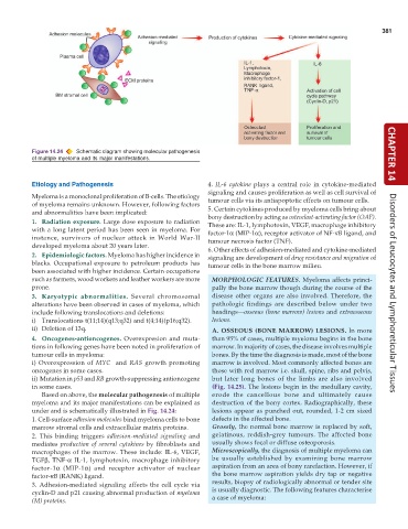
           Figure 14.24  Schematic diagram showing molecular pathogenesis                                             CHAPTER 14
           of multiple myeloma and its major manifestations.


           Etiology and Pathogenesis                           4. IL-6 cytokine plays a central role in cytokine-mediated
                                                               signaling and causes proliferation as well as cell survival of
           Myeloma is a monoclonal proliferation of B-cells. The etiology  tumour cells via its antiapoptotic effects on tumour cells.
           of myeloma remains unknown. However, following factors
           and abnormalities have been implicated:             5. Certain cytokines produced by myeloma cells bring about
                                                               bony destruction by acting as osteoclast-activating factor (OAF).
           1. Radiation exposure. Large dose exposure to radiation  These are: IL-1, lymphotoxin, VEGF, macrophage inhibitory
           with a long latent period has been seen in myeloma. For  factor-1α (MIP-1α), receptor activator of NF-κB ligand, and
           instance, survivors of nuclear attack in World War-II  tumour necrosis factor (TNF).
           developed myeloma about 20 years later.             6. Other effects of adhesion-mediated and cytokine-mediated
           2. Epidemiologic factors. Myeloma has higher incidence in  signaling are development of drug resistance and migration of
           blacks. Occupational exposure to petroleum products has  tumour cells in the bone marrow milieu.
           been associated with higher incidence. Certain occupations
           such as farmers, wood workers and leather workers are more  MORPHOLOGIC FEATURES. Myeloma affects princi-
           prone.                                                pally the bone marrow though during the course of the
           3. Karyotypic abnormalities.  Several chromosomal     disease other organs are also involved. Therefore, the  Disorders of Leucocytes and Lymphoreticular Tissues
           alterations have been observed in cases of myeloma, which  pathologic findings are described below under two
           include following translocations and deletions:       headings—osseous (bone marrow) lesions and extraosseous
           i) Translocations t(11;14)(q13;q32) and t(4;14)(p16;q32).  lesions.
           ii) Deletion of 13q.                                  A. OSSEOUS (BONE MARROW) LESIONS. In more
           4. Oncogenes-antioncogenes. Overexpresion and muta-   than 95% of cases, multiple myeloma begins in the bone
           tions in following genes have been noted in proliferation of  marrow. In majority of cases, the disease involves multiple
           tumour cells in myeloma:                              bones. By the time the diagnosis is made, most of the bone
           i) Overexpression of  MYC  and  RAS  growth promoting  marrow is involved. Most commonly affected bones are
           oncogenes in some cases.                              those with red marrow i.e. skull, spine, ribs and pelvis,
           ii)  Mutation in p53 and RB growth-suppressing antioncogene  but later long bones of the limbs are also involved
           in some cases.                                        (Fig. 14.25). The lesions begin in the medullary cavity,
              Based on above, the molecular pathogenesis of multiple  erode the cancellous bone and ultimately cause
           myeloma and its major manifestations can be explained as  destruction of the bony cortex. Radiographically, these
           under and is schematically illustrated in Fig. 14.24:  lesions appear as punched out, rounded, 1-2 cm sized
           1. Cell-surface adhesion molecules bind myeloma cells to bone  defects in the affected bone.
           marrow stromal cells and extracellular matrix proteins.  Grossly, the normal bone marrow is replaced by soft,
           2. This binding triggers  adhesion-mediated signaling and  gelatinous, reddish-grey tumours. The affected bone
           mediates  production of several cytokines by fibroblasts and  usually shows focal or diffuse osteoporosis.
           macrophages of the marrow. These include: IL-6, VEGF,  Microscopically, the diagnosis of multiple myeloma can
           TGFβ, TNF-α IL-1, lymphotoxin, macrophage inhibitory  be usually established by examining bone marrow
           factor-1α (MIP-1α) and receptor activator of nuclear  aspiration from an area of bony rarefaction. However, if
           factor-κB (RANK) ligand.                              the bone marrow aspiration yields dry tap or negative
           3. Adhesion-mediated signaling affects the cell cycle via  results, biopsy of radiologically abnormal or tender site
                                                                 is usually diagnostic. The following features characterise
           cyclin-D and p21 causing abnormal production of myeloma  a case of myeloma:
           (M) proteins.

