


Page 402 - Textbook of Pathology, 6th Edition
P. 402
386                                                      cases. The condition is currently regarded as an unusual form
                                                               of malignant lymphoma.

                                                                                    SPLEEN
                                                               NORMAL STRUCTURE

                                                               The spleen is the largest lymphoid organ of the body. Under
                                                               normal conditions, the average weight of the spleen is about
                                                               150 gm in the adult. Normally, the organ lies well protected
                                                               by the 9th, 10th and 11th ribs in the upper left quadrant. The
                                                               surface of the spleen is covered by a layer of peritoneum
                                                               underneath which the organ is ensheathed by a thin capsule.
                                                               From the capsule extend connective tissue trabeculae into the
                                                               pulp of the organ and serve as supportive network. Blood
                                                               enters the spleen by the splenic artery which divides into
                                                               branches that penetrate the spleen via trabeculae. From the
                                                               trabeculae arise small branches called central arterioles. Blood
                                                               in the central arterioles empties partly into splenic venules
                                                               and from there into splenic vein, but largely into vascular
     SECTION II
                                                               sinuses of the red pulp and thence into the splenic venous
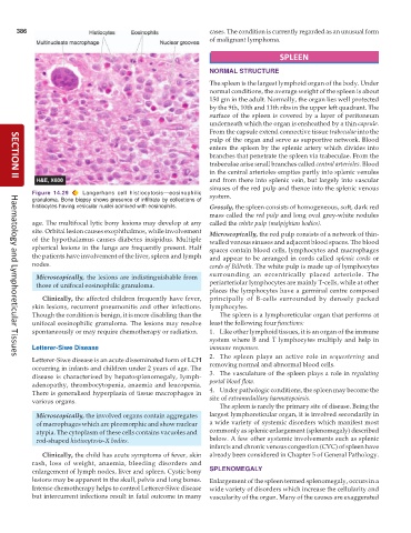
           Figure 14.29   Langerhans cell histiocytosis—eosinophilic  system.
           granuloma. Bone biopsy shows presence of infiltrate by collections of
           histiocytes having vesicular nuclei admixed with eosinophils.  Grossly, the spleen consists of homogeneous, soft, dark red
                                                               mass called the red pulp and long oval grey-white nodules
           age. The multifocal lytic bony lesions may develop at any  called the white pulp (malpighian bodies).
           site. Orbital lesion causes exophthalmos, while involvement  Microscopically, the red pulp consists of a network of thin-
           of the hypothalamus causes diabetes insipidus. Multiple  walled venous sinuses and adjacent blood spaces. The blood
           spherical lesions in the lungs are frequently present. Half  spaces contain blood cells, lymphocytes and macrophages
           the patients have involvement of the liver, spleen and lymph  and appear to be arranged in cords called splenic cords or
           nodes.                                              cords of Billroth. The white pulp is made up of lymphocytes

            Microscopically, the lesions are indistinguishable from  surrounding an eccentrically placed arteriole. The
            those of unifocal eosinophilic granuloma.          periarteriolar lymphocytes are mainly T-cells, while at other
                                                               places the lymphocytes have a germinal centre composed
              Clinically, the affected children frequently have fever,  principally of B-cells surrounded by densely packed
           skin lesions, recurrent pneumonitis and other infections.  lymphocytes.
           Though the condition is benign, it is more disabling than the  The spleen is a lymphoreticular organ that performs at
           unifocal eosinophilic granuloma. The lesions may resolve  least the following four functions:
           spontaneously or may require chemotherapy or radiation.  1. Like other lymphoid tissues, it is an organ of the immune
                                                               system where B and T lymphocytes multiply and help in
           Letterer-Siwe Disease                               immune responses.
                                                               2. The spleen plays an active role in  sequestering  and
           Letterer-Siwe disease is an acute disseminated form of LCH
     Haematology and Lymphoreticular Tissues
           occurring in infants and children under 2 years of age. The  removing normal and abnormal blood cells.
           disease is characterised by hepatosplenomegaly, lymph-  3. The vasculature of the spleen plays a role in regulating
           adenopathy, thrombocytopenia, anaemia and leucopenia.  portal blood flow.
           There is generalised hyperplasia of tissue macrophages in  4. Under pathologic conditions, the spleen may become the
           various organs.                                     site of extramedullary haematopoiesis.
                                                                  The spleen is rarely the primary site of disease. Being the
            Microscopically, the involved organs contain aggregates  largest lymphoreticular organ, it is involved secondarily in
            of macrophages which are pleomorphic and show nuclear  a wide variety of systemic disorders which manifest most
            atypia. The cytoplasm of these cells contains vacuoles and  commonly as splenic enlargement (splenomegaly) described
            rod-shaped histiocytosis-X bodies.                 below. A few other systemic involvements such as splenic
                                                               infarcts and chronic venous congestion (CVC) of spleen have
              Clinically, the child has acute symptoms of fever, skin  already been considered in Chapter 5 of General Pathology.
           rash, loss of weight, anaemia, bleeding disorders and
           enlargement of lymph nodes, liver and spleen. Cystic bony  SPLENOMEGALY
           lesions may be apparent in the skull, pelvis and long bones.  Enlargement of the spleen termed splenomegaly, occurs in a
           Intense chemotherapy helps to control Letterer-Siwe disease  wide variety of disorders which increase the cellularity and
           but intercurrent infections result in fatal outcome in many  vascularity of the organ. Many of the causes are exaggerated

