


Page 415 - Textbook of Pathology, 6th Edition
P. 415
399


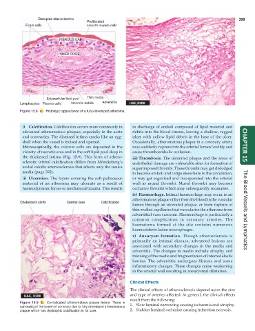
           Figure 15.8  Histologic appearance of a fully-developed atheroma.


            i) Calcification. Calcification occurs more commonly in  in discharge of emboli composed of lipid material and
            advanced atheromatous plaques, especially in the aorta  debris into the blood stream, leaving a shallow, ragged
            and coronaries. The diseased intima cracks like an egg-  ulcer with yellow lipid debris in the base of the ulcer.
            shell when the vessel is incised and opened.         Occasionally, atheromatous plaque in a coronary artery
            Microscopically, the calcium salts are deposited in the  may suddenly rupture into the arterial lumen forcibly and  CHAPTER 15
            vicinity of necrotic area and in the soft lipid pool deep in  cause thromboembolic occlusion.
            the thickened intima  (Fig. 15.9). This form of athero-  iii) Thrombosis. The ulcerated plaque and the areas of
            sclerotic intimal calcification differs from Mönckeberg’s  endothelial damage are vulnerable sites for formation of
            medial calcific arteriosclerosis that affects only the tunica  superimposed thrombi. These thrombi may get dislodged
            media (page 392).                                    to become emboli and lodge elsewhere in the circulation,
            ii) Ulceration. The layers covering the soft pultaceous  or may get organised and incorporated into the arterial
            material of an atheroma may ulcerate as a result of  wall as mural thrombi. Mural thrombi may become
            haemodynamic forces or mechanical trauma. This results  occlusive thrombi which may subsequently recanalise.
                                                                 iv) Haemorrhage. Intimal haemorrhage may occur in an
                                                                 atheromatous plaque either from the blood in the vascular
                                                                 lumen through an ulcerated plaque, or from rupture of
                                                                 thin-walled capillaries that vascularise the atheroma from  The Blood Vessels and Lymphatics
                                                                 adventitial vasa vasorum. Haemorrhage is particularly a
                                                                 common complication in coronary arteries. The
                                                                 haematoma formed at the site contains numerous
                                                                 haemosiderin-laden macrophages.
                                                                 v) Aneurysm formation. Though atherosclerosis is
                                                                 primarily an intimal disease, advanced lesions are
                                                                 associated with secondary changes in the media and
                                                                 adventitia. The changes in media include atrophy and
                                                                 thinning of the media and fragmentation of internal elastic
                                                                 lamina. The adventitia undergoes fibrosis and some
                                                                 inflammatory changes. These changes cause weakening
                                                                 in the arterial wall resulting in aneurysmal dilatation.


                                                               Clinical Effects
                                                               The clinical effects of atherosclerosis depend upon the size
                                                               and type of arteries affected. In general, the clinical effects
                                                               result from the following:
           Figure 15.9  Complicated atheromatous plaque lesion. There is  1. Slow luminal narrowing causing ischaemia and atrophy.
           narrowing of the lumen of coronary due to fully developed atheromatous
           plaque which has dystrophic calcification in its core.  2. Sudden luminal occlusion causing infarction necrosis.

