


Page 412 - Textbook of Pathology, 6th Edition
P. 412
396                                                         In man, two of the major risk factors which act together
                                                               to produce endothelial injury are: haemodynamic stress from
                                                               hypertension and chronic dyslipidaemia. The role of
                                                               haemodynamic forces in causing endothelial injury is further
                                                               supported by the distribution of atheromatous plaques at
                                                               points of bifurcation or branching of blood vessels which are
                                                               under greatest shear stress.
                                                               ii) Intimal smooth muscle cell proliferation. Endothelial
                                                               injury causes adherence, aggregation and platelet release
                                                               reaction at the site of exposed subendothelial connective
                                                               tissue and infiltration by inflammatory cells. Proliferation of
                                                               intimal smooth muscle cell and production of extracellular
                                                               matrix are stimulated by various cytokines such as IL-1 and
                                                               TNF-α released from invading monocyte-macrophages and
                                                               by activated platelets at the site of endothelial injury. These
                                                               cytokines lead to local synthesis of following growth factors
                                                               having distinct roles in plaque evolution:
                                                                  Platelet-derived growth factor (PDGF) and fibroblast
                                                               growth factor (FGF) stimulate proliferation and migration
                                                               of smooth muscle cells from their usual location in the media
                                                               into the intima.
                                                                  Transforming growth factor-β (TGF-β)  and interferon-
                                                               (IFN-γ) derived from activated T lymphocytes within lesions
                                                               regulate the synthesis of collagen by smooth muscle cells.
                                                                  Smooth muscle cell proliferation is also facilitated by
                                                               biomolecules such as nitric oxide and endothelin released from
                                                               endothelial cells. Intimal proliferation of smooth muscle cells
                                                               is accompanied by synthesis of matrix proteins—collagen,
     SECTION III
                                                               elastic fibre proteins and proteoglycans.
                                                               iii) Role of blood monocytes. Though blood monocytes do
                                                               not possess receptors for normal LDL, LDL does appear in
                                                               the monocyte cytoplasm to form foam cell by mechanism
                                                               illustrated in Fig. 15.5. Plasma LDL on entry into the intima
                                                               undergoes oxidation. The ‘oxidised LDL’ formed in the
                                                               intima performs the following all-important functions on
                                                               monocytes and endothelium:
                                                                  For monocytes: Oxidised LDL acts to attract, proliferate,
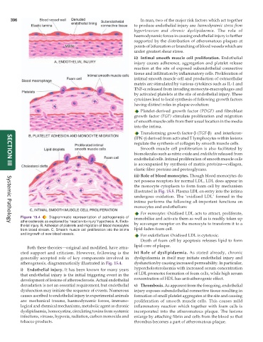
           Figure 15.4  Diagrammatic representation of pathogenesis of  immobilise and activate them as well as is readily taken up
           atherosclerosis as explained by ‘reaction-to-injury’ hypothesis. A, Endo-  by scavenger receptor on the monocyte to transform it to a
     Systemic Pathology
           thelial injury. B, Adhesion of platelets and migration of blood monocytes
           from blood stream. C, Smooth muscle cell proliferation into the intima  lipid-laden foam cell.
           and ingrowth of new blood vessels.                     For endothelium: Oxidised LDL is cytotoxic.
                                                                  Death of foam cell by apoptosis releases lipid to form
              Both these theories—original and modified, have attra-  lipid core of plaque.
           cted support and criticism. However, following is the  iv) Role of dyslipidaemia. As stated already, chronic
           generally accepted role of key components involved in  dyslipidaemia in itself may initiate endothelial injury and
           atherogenesis, diagrammatically illustrated in Fig. 15.4.  dysfunction by causing increased permeability. In particular,
           i) Endothelial injury. It has been known for many years  hypercholesterolaemia with increased serum concentration
           that endothelial injury is the initial triggering event in the  of LDL promotes formation of foam cells, while high serum
           development of lesions of atherosclerosis. Actual endothelial  concentration of HDL has anti-atherogenic effect.
           denudation is not an essential requirement, but endothelial  v) Thrombosis. As apparent from the foregoing, endothelial
           dysfunction may initiate the sequence of events. Numerous  injury exposes subendothelial connective tissue resulting in
           causes ascribed to endothelial injury in experimental animals  formation of small platelet aggregates at the site and causing
           are: mechanical trauma, haemodynamic forces, immuno-  proliferation of smooth muscle cells. This causes mild
           logical and chemical mechanisms, metabolic agent as chronic  inflammatory reaction which together with foam cells is
           dyslipidaemia, homocystine, circulating toxins from systemic  incorporated into the atheromatous plaque. The lesions
           infections, viruses, hypoxia, radiation, carbon monoxide and  enlarge by attaching fibrin and cells from the blood so that
           tobacco products.                                   thrombus becomes a part of atheromatous plaque.

