


Page 414 - Textbook of Pathology, 6th Edition
P. 414
398


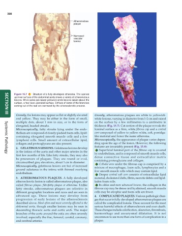
           Figure 15.7  Structure of a fully-developed atheroma. The opened
           up inner surface of the abdominal aorta shows a variety of atheromatous
           lesions. While some are raised yellowish-white lesions raised above the
           surface, a few have ulcerated surface. Orifices of some of the branches
           coming out of the wall are narrowed by the atherosclerotic process.


            Grossly, the lesions may appear as flat or slightly elevated  Grossly, atheromatous plaques are white to yellowish-
     SECTION III
            and yellow. They may be either in the form of small,  white lesions, varying in diameter from 1-2 cm and raised
            multiple dots, about 1 mm in size, or in the form of  on the surface by a few millimetres to a centimetre in
            elongated, beaded streaks.                           thickness (Fig. 15.7). Cut section of the plaque reveals the
            Microscopically, fatty streaks lying under the endo-  luminal surface as a firm, white fibrous cap and a central
            thelium are composed of closely-packed foam cells, lipid-  core composed of yellow to yellow-white, soft, porridge-
            containing elongated smooth muscle cells and a few   like material and hence the name atheroma.
            lymphoid cells. Small amount of extracellular lipid,  Microscopically, the appearance of plaque varies depen-
            collagen and proteoglycans are also present.         ding upon the age of the lesion. However, the following
                                                                 features are invariably present (Fig. 15.8):
            2. GELATINOUS LESIONS. Gelatinous lesions develop
                                                                    Superficial luminal part of the fibrous cap is covered
            in the intima of the aorta and other major arteries in the  by endothelium, and is composed of smooth muscle cells,
            first few months of life. Like fatty streaks, they may also  dense connective tissue and extracellular matrix
     Systemic Pathology
            be precursors of plaques. They are round or oval,    containing proteoglycans and collagen.
            circumscribed grey elevations, about 1 cm in diameter.  Cellular area under the fibrous cap is comprised by a
            Microscopically, gelatinous lesions are foci of increased  mixture of macrophages, foam cells, lymphocytes and a
            ground substance in the intima with thinned overlying  few smooth muscle cells which may contain lipid.
            endothelium.                                            Deeper central soft core consists of extracellular lipid
            3. ATHEROMATOUS PLAQUES. A fully developed           material, cholesterol clefts, fibrin, necrotic debris and lipid-
            atherosclerotic lesion is called atheromatous plaque, also  laden foam cells.
            called fibrous plaque, fibrofatty plaque or atheroma. Unlike  In older and more advanced lesions, the collagen in the
            fatty streaks, atheromatous plaques are selective in  fibrous cap may be dense and hyalinised, smooth muscle
            different geographic locations and races and are seen in  cells may be atrophic and foam cells are fewer.
            advanced age. These lesions may develop from         4. COMPLICATED PLAQUES. Various pathologic chan-
            progression of early lesions of the atherosclerosis  ges that occur in fully-developed atheromatous plaques are
            described above. Most often and most severely affected is the  called the complicated lesions. These account for the most
            abdominal aorta, though smaller lesions may be seen in  serious harmful effects of atherosclerosis and even death.
            descending thoracic aorta and aortic arch. The major  These changes include calcification, ulceration, thrombosis,
            branches of the aorta around the ostia are often severely  haemorrhage and aneurysmal dilatation. It is not
            involved, especially the iliac, femoral, carotid, coronary,  uncommon to see more than one form of complication in a
            and cerebral arteries.                               plaque.

