


Page 413 - Textbook of Pathology, 6th Edition
P. 413
397


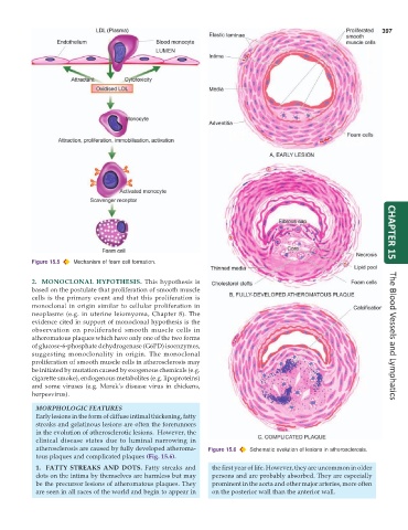
           Figure 15.5  Mechanism of foam cell formation.                                                             CHAPTER 15


           2. MONOCLONAL HYPOTHESIS.  This hypothesis is
           based on the postulate that proliferation of smooth muscle
           cells is the primary event and that this proliferation is
           monoclonal in origin similar to cellular proliferation in
           neoplasms (e.g. in uterine leiomyoma, Chapter 8). The
           evidence cited in support of monoclonal hypothesis is the
           observation on proliferated smooth muscle cells in
           atheromatous plaques which have only one of the two forms                                                  The Blood Vessels and Lymphatics
           of glucose-6-phosphate dehydrogenase (G6PD) isoenzymes,
           suggesting monoclonality in origin. The monoclonal
           proliferation of smooth muscle cells in atherosclerosis may
           be initiated by mutation caused by exogenous chemicals (e.g.
           cigarette smoke), endogenous metabolites (e.g. lipoproteins)
           and some viruses (e.g. Marek’s disease virus in chickens,
           herpesvirus).

            MORPHOLOGIC FEATURES
            Early lesions in the form of diffuse intimal thickening, fatty
            streaks and gelatinous lesions are often the forerunners
            in the evolution of atherosclerotic lesions.  However, the
            clinical disease states due to luminal narrowing in
            atherosclerosis are caused by fully developed atheroma-  Figure 15.6  Schematic evolution of lesions in atherosclerosis.
            tous plaques and complicated plaques (Fig. 15.6).

            1. FATTY STREAKS AND DOTS. Fatty streaks and         the first year of life. However, they are uncommon in older
            dots on the intima by themselves are harmless but may  persons and are probably absorbed. They are especially
            be the precursor lesions of atheromatous plaques. They  prominent in the aorta and other major arteries, more often
            are seen in all races of the world and begin to appear in  on the posterior wall than the anterior wall.

