


Page 433 - Textbook of Pathology, 6th Edition
P. 433
417


            Chapter 16
            Chapter 16                                         The Heart


                       NORMAL STRUCTURE                           Wall of the heart consists mainly of the myocardium which
                                                               is covered externally by thin membrane, the epicardium or
           ANATOMY AND PHYSIOLOGY. The heart is a muscular     visceral pericardium, and lined internally by another thin
           pump that ejects blood into the vascular tree with sufficient  layer, the endocardium.
           pressure to maintain optimal circulation. Average weight of  The  myocardium is the muscle tissue of the heart
           the heart in an adult male is 300-350 gm while that of an  composed of syncytium of branching and anastomosing,
           adult female is 250-300 gm. Heart is divided into four  transversely striated muscle fibres arranged in parallel
           chambers: a right and a left atrium both lying superiorly,  fashion. The space between myocardial fibres contains a rich
           and a right and a left ventricle both lying inferiorly and are  capillary network and loose connective tissue. The
           larger. The atria are separated by a thin interatrial partition  myocardial fibres are connected to each other by irregular
           called interatrial septum, while the ventricles are separated  joints called as intercalated discs. They represent apposed cell
           by thick muscular partition called interventricular septum. The  membranes of individual cells which act as tight junctions
           thickness of the right ventricular wall is 0.3 to 0.5 cm while  for free transport of ions and action potentials. The cardiac
           that of the left ventricular wall is 1.3 to 1.5 cm. The blood in  myocyte is very rich in mitochondria which is the source of
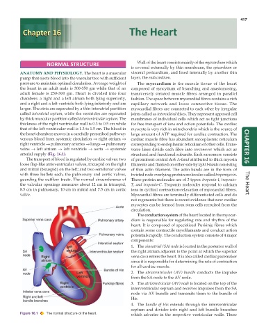
           the heart chambers moves in a carefully prescribed pathway:  large amount of ATP required for cardiac contraction. The
           venous blood from systemic circulation → right atrium →  cardiac muscle fibre has abundant sarcoplasmic reticulum
           right ventricle → pulmonary arteries → lungs → pulmonary  corresponding to endoplasmic reticulum of other cells. Trans-
           veins  → left atrium → left ventricle → aorta  → systemic  verse lines divide each fibre into sarcomeres which act as  CHAPTER 16
           arterial supply (Fig. 16.1).                        structural and functional subunits. Each sarcomere consists
              The transport of blood is regulated by cardiac valves: two  of prominent central dark A-band attributed to thick myosin
           loose flap-like atrioventricular valves, tricuspid on the right  filaments and flanked on either side by light I-bands consisting
           and mitral (bicuspid) on the left; and two semilunar valves  of thin actin filament. The actin bands are in the form of
           with three leaflets each, the pulmonary and aortic valves,  twisted rods overlying protein molecules called tropomyosin.
           guarding the outflow tracts. The normal circumference of  These protein molecules are of 3 types: troponin-I, troponin-
           the valvular openings measures about 12 cm in tricuspid,  T, and troponin-C. Troponin molecules respond to calcium  The Heart
           8.5 cm in pulmonary, 10 cm in mitral and 7.5 cm in aortic  ions in cyclical contraction-relaxation of myocardial fibres.
           valve.                                              Myocardial fibres are terminally differentiated cells and do
                                                               not regenerate but there is recent evidence that new cardiac
                                                               myocytes can be formed from stem cells recruited from the
                                                               circulation.
                                                                  The conduction system of the heart located in the myocar-
                                                               dium is responsible for regulating rate and rhythm of the
                                                               heart. It is composed of specialised Purkinje fibres which
                                                               contain some contractile myofilaments and conduct action
                                                               potentials rapidly. The conduction system consists of 4 major
                                                               components:
                                                               1. The sinoatrial (SA) node is located in the posterior wall of
                                                               the right atrium adjacent to the point at which the superior
                                                               vena cava enters the heart. It is also called cardiac pacemaker
                                                               since it is responsible for determining the rate of contraction
                                                               for all cardiac muscle.
                                                               2. The  atrioventricular (AV) bundle conducts the impulse
                                                               from the SA node to the AV node.
                                                               3. The atrioventricular (AV) node is located on the top of the
                                                               interventricular septum and receives impulses from the SA
                                                               node via AV bundle and transmits them to the bundle of
                                                               His.
                                                               4. The  bundle of His extends through the interventricular
                                                               septum and divides into right and left bundle branches
           Figure 16.1  The normal structure of the heart.     which arborise in the respective ventricular walls. These

