


Page 468 - Textbook of Pathology, 6th Edition
P. 468
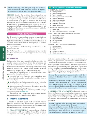
452 Microscopically, the enlarged cusp shows loose TABLE 16.9: Etiologic Classification of Myocarditis.
connective tissue with abundant mucoid or myxoid I. INFECTIVE MYOCARDITIS
material due to abundance of mucopolysaccharide. 1. Viral myocarditis
2. Suppurative myocarditis
EFFECTS. Usually the condition does not produce any 3. Toxic myocarditis
symptoms or significant valvular dysfunction. The condition 4. Infective granulomatous myocarditis
is recognised during life by the characteristic mid-systolic 5. Syphilitic myocarditis
click followed by a systolic murmur due to mildly 6. Rickettsial myocarditis
incompetent mitral valve caused by the mitral valve prolapse. 7. Protozoal myocarditis
Occasionally, complications may develop such as 8. Helminthic myocarditis
superimposed infective endocarditis, mitral insufficiency and 9. Fungal myocarditis
arrhythmias. Rarely, sudden death from serious ventricular II. IDIOPATHIC (FIEDLER’S) MYOCARDITIS
arrhythmias may occur. 1. Diffuse type
2. Giant cell (idiopathic granulomatous) type
MYOCARDIAL DISEASE III. MYOCARDITIS IN CONNECTIVE TISSUE DISEASES
1. Rheumatoid arthritis
Involvement of the myocardium occurs in three major forms 2. Lupus erythematosus
of diseases already discussed—ischaemic heart disease, 3. Polyarteritis nodosa
hypertensive heart disease and rheumatic heart disease. In 4. Dermatomyositis
addition, there are two other broad groups of isolated 5. Scleroderma
myocardial diseases: IV. MISCELLANEOUS TYPES OF MYOCARDITIS
I. Myocarditis i.e. inflammatory involvement of the 1. Physical agents
myocardium; and 2. Chemical agents
3. Drugs
II. Cardiomyopathy i.e. a non-inflammatory myocardial 4. Immunologic agents
involvement with unknown (primary) or known (secondary) 5. Metabolic derangements
etiology.
MYOCARDITIS nucleosis, hepatitis, smallpox, chickenpox, measles, mumps,
SECTION III
Inflammation of the heart muscle is called myocarditis. It is rubella, viral pneumonias, coxsackievirus and HIV infections.
a rather common form of heart disease that can occur at any Cardiac involvement occurs in about 5% of viral infections.
age. Its exact incidence is difficult to ascertain as the Viral myocarditis usually appears after a few days to a few
histological examination has been largely confined to autopsy weeks of viral infections elsewhere in the body. The damage
material. Reports from different studies have estimated the to the myocardium is caused either by direct viral cytotoxi-
incidence of myocarditis in 1 to 4% of all autopsies. city or by cell-mediated immune reaction. Regardless of the
A number of classifications of myocarditis have been type of virus, the pathologic changes are similar.
proposed in the past as follows: Grossly, the myocardium is pale and flabby with dila-
Interstitial and parenchymatous type, depending upon tation of the chambers. There may be focal or patchy areas
whether the inflammation is confined to interstitial tissue or of necrosis.
the parenchyma. Histologically, there are changes of acute myocarditis.
Systemic Pathology
Specific and non-specific type, depending upon whether the Initially, there is oedema and infiltration of the interstitial
inflammation is granulomatous or non-specific type. tissue by neutrophils and lymphocytes. Later, there is
Acute, subacute and chronic type, depending upon the necrosis of individual myocardial fibres and the infiltrate
duration of inflammatory response. consists of lymphocytes and macrophages.
However, currently most commonly used is etiologic
classification given in Table 16.9. 2. SUPPURATIVE MYOCARDITIS. Pyogenic bacteria,
According to this classification, myocarditis is divided chiefly Staphylococcus aureus or Streptococcus pyogenes, which
into 4 main etiologic types described below. cause septicaemia and pyaemia may produce suppurative
myocarditis. As already pointed out, acute bacterial
I. INFECTIVE MYOCARDITIS endocarditis may sometimes cause bacterial myocarditis
(page 445).
A number of infectious agents such as bacteria, viruses,
protozoa, parasites, fungi, rickettsiae and spirochaetes may Grossly, There are either abscesses in the myocardium
cause myocarditis by direct invasion or by their toxins. Some or there is diffuse myocardial involvement.
of the common forms are described below. Microscopically, the exudate chiefly consists of neutro-
phils, admixed with lymphocytes, plasma cells and
1. VIRAL MYOCARDITIS. A number of viral infections macrophages. There may be foci of myocardial degene-
are associated with myocarditis. Some of the common ration and necrosis with areas of healing by fibrosis.
examples are influenza, poliomyelitis, infectious mono-

