


Page 465 - Textbook of Pathology, 6th Edition
P. 465
capsulatum, Aspergillus, Mucor, coccidioidomycosis, crypto-  The major forms of vegetative endocarditis involving the  449
           coccosis, blastomycosis and actinomycosis. Opportunistic  valves have already been described. Others along with the
           fungal infections like candidiasis and aspergillosis are seen  consequences of these valvular diseases in the form of
           more commonly in patients receiving long-term antibiotic  stenosis and insufficiency of the heart valves are described
           therapy, intravenous drug abusers and after prosthetic valve  below.
           replacement. Fungal endocarditis produces appearance
           similar to that in BE but the vegetations are bulkier in fungal  MITRAL STENOSIS
           endocarditis.
                                                               Mitral stenosis occurs in approximately 40% of all patients
           4. Viral endocarditis. There is only experimental evidence  with RHD. About 70% of the patients are women. The latent
           of existence of this entity.                        period between the rheumatic carditis and development of
           5. Rickettsial endocarditis. Another rare cause of  symptomatic mitral stenosis is about two decades.
           endocarditis is from infection with rickettsiae in Q fever.
                                                               ETIOLOGY. Mitral stenosis is generally rheumatic in origin.
                                                               Less common causes include bacterial endocarditis, Libman-
             VALVULAR DISEASES AND DEFORMITIES
                                                               Sacks endocarditis, endocardial fibroelastosis and congenital
           Valvular diseases are various forms of congenital and  parachute mitral valve.
           acquired diseases which cause valvular deformities. Many
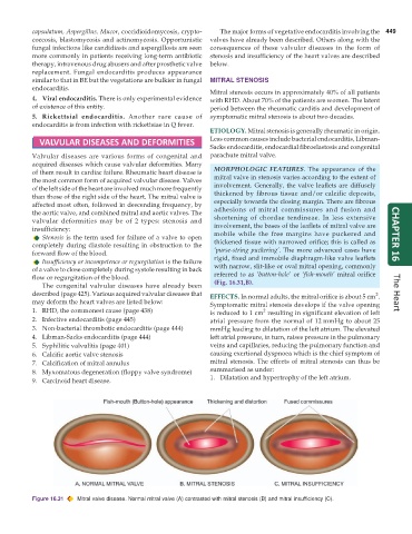
           of them result in cardiac failure. Rheumatic heart disease is  MORPHOLOGIC FEATURES. The appearance of the
           the most common form of acquired valvular disease. Valves  mitral valve in stenosis varies according to the extent of
           of the left side of the heart are involved much more frequently  involvement. Generally, the valve leaflets are diffusely
           than those of the right side of the heart. The mitral valve is  thickened by fibrous tissue and/or calcific deposits,
           affected most often, followed in descending frequency, by  especially towards the closing margin. There are fibrous
           the aortic valve, and combined mitral and aortic valves. The  adhesions of mitral commissures and fusion and
           valvular deformities may be of 2 types: stenosis and  shortening of chordae tendineae. In less extensive
           insufficiency:                                        involvement, the bases of the leaflets of mitral valve are
              Stenosis is the term used for failure of a valve to open  mobile while the free margins have puckered and  CHAPTER 16
           completely during diastole resulting in obstruction to the  thickened tissue with narrowed orifice; this is called as
           forward flow of the blood.                            ‘purse-string puckering’. The more advanced cases have
              Insufficiency or incompetence or regurgitation is the failure  rigid, fixed and immobile diaphragm-like valve leaflets
           of a valve to close completely during systole resulting in back  with narrow, slit-like or oval mitral opening, commonly
           flow or regurgitation of the blood.                   referred to as ‘button-hole’ or ‘fish-mouth’ mitral orifice
              The congenital valvular diseases have already been  (Fig. 16.31,B).
           described (page 425). Various acquired valvular diseases that  EFFECTS. In normal adults, the mitral orifice is about 5 cm .  The Heart
                                                                                                                 2
           may deform the heart valves are listed below:       Symptomatic mitral stenosis develops if the valve opening
           1. RHD, the commonest cause (page 438)              is reduced to 1 cm  resulting in significant elevation of left
                                                                               2
           2. Infective endocarditis (page 445)                atrial pressure from the normal of 12 mmHg to about 25
           3. Non-bacterial thrombotic endocarditis (page 444)  mmHg leading to dilatation of the left atrium. The elevated
           4. Libman-Sacks endocarditis (page 444)             left atrial pressure, in turn, raises pressure in the pulmonary
           5. Syphilitic valvulitis (page 401)                 veins and capillaries, reducing the pulmonary function and
           6. Calcific aortic valve stenosis                   causing exertional dyspnoea which is the chief symptom of
           7. Calcification of mitral annulus                  mitral stenosis. The effects of mitral stenosis can thus be
           8. Myxomatous degeneration (floppy valve syndrome)  summarised as under:
           9. Carcinoid heart disease.                         1. Dilatation and hypertrophy of the left atrium.


           Figure 16.31  Mitral valve disease. Normal mitral valve (A) contrasted with mitral stenosis (B) and mitral insufficiency (C).

