


Page 463 - Textbook of Pathology, 6th Edition
P. 463
447
TABLE 16.8: Distinguishing Features of Vegetations in Major Forms of Endocarditis.
Feature Rheumatic Libman-Sacks Non-bacterial Bacterial
Thrombotic
1. Valves commonly Mitral alone; mitral Mitral, tricuspid Mainly mitral; less often Mitral; aortic; combined
affected and aortic combined aortic and tricuspid mitral and aortic
2. Location on valve Occur along the line of Occur on both sur- Occur along the line of SABE more often on
cusps or leaflets closure, atrial surface of faces of valve leaflets closure diseased valves: ABE on
atrioventricular valves or cusps, in the valve previously normal valves;
and ventricular surface pockets location same as in RHD
of semilunar valves
3. Macroscopy Small, multiple, warty, Medium-sized, multi- Small but larger than Often large, grey-tawny
grey brown, translucent, ple, generally do not those of rheumatic, to greenish, irregular,
firmly attached, generally produce significant single or multiple, single or multiple,
produce permanent val- valvular deformity brownish, firm, but typically friable
vular deformity more friable than those
of rheumatic
4. Microscopy Composed of fibrin with Composed of fibrinoid Composed of degenerated Composed of outer
superimposed platelet material with superim- valvular tissue, fibrin- eosinophilic zone of fibrin
thrombi and no bacteria, posed fibrin and platelet platelets thrombi and no and platelets, covering
Adjacent and underlying thrombi and no bacteria. bacteria. The underlying colonies of bacteria and
endocardium shows The underlying endo- valve shows swelling of deeper zone of non-specific
oedema, proliferation of cardium shows fibrinoid collagen, fibrinoid change, acute and chronic inflam-
capillaries, mononuclear necrosis, proliferation of proliferation of capillaries matory cells. The underlying
inflammatory infiltrate capillaries and acute and but no significant inflam- endocardium may show
and occasional Aschoff chronic inflammatory matory cell infiltrate. abscesses in ABE and
bodies. infiltrate including the inflammatory granulation
haematoxylin bodies tissue in the SABE.
of Gross. CHAPTER 16
accompanied with tissue necrosis and abscesses in the valve COMPLICATIONS AND SEQUELAE. Most cases of BE
rings and in the myocardium. In the subacute form, there is present with fever. The acute form of BE is characterised by
healing by granulation tissue, mononuclear inflammatory cell high grade fever, chills, weakness and malaise while the
infiltration and fibroblastic proliferation. Histological subacute form of the disease has non-specific manifestations
evidence of pre-existing valvular disease such as RHD may like slight fever, fatigue, loss of weight and flu-like The Heart
be present in SABE. symptoms. In the early stage, the lesions are confined to the
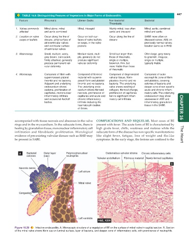
Figure 16.29 Infective endocarditis. A, Microscopic structure of a vegetation of BE on the surface of mitral valve in sagittal section. B, Section
of the mitral valve shows fibrin cap on luminal surface, layer of bacteria, and deeper zone of inflammatory cells, with prominence of neutrophils.

