


Page 466 - Textbook of Pathology, 6th Edition
P. 466
450 2. Normal-sized or atrophic left ventricle due to reduced  as well as pressure since the left ventricle cannot empty
           inflow of blood.                                    completely. This results in rise in left atrial pressure and
           3. Pulmonary hypertension resulting from passive    dilatation. As a consequence of left atrial hypertension,
           backward transmission of elevated left artial pressure which  pulmonary hypertension occurs resulting in pulmonary
           causes:                                             oedema and right heart failure. In symptomatic cases of
            i) chronic passive congestion of the lungs;        mitral insufficiency, the major symptoms are related to
            ii) hypertrophy and dilatation of the right ventricle; and  decreased cardiac output (e.g. fatigue and weakness) and
           iii) dilatation of the right atrium when right heart failure  due to pulmonary congestion (e.g. exertional dyspnoea and
               supervenes.                                     orthopnoea) but the features are less well marked than in
                                                               mitral stenosis. The effects of mitral insufficiency may be
           MITRAL INSUFFICIENCY                                summarised as under:
                                                               1. Dilatation and hypertrophy of the left ventricle.
           Mitral insufficiency is caused by RHD in about 50% of  2. Marked dilatation of the left atrium.
           patients but in contrast to mitral stenosis, pure mitral  3. Features of pulmonary hypertension such as:
           insufficiency occurs more often in men (75%). Subsequently,  i) chronic passive congestion of the lungs;
           mitral insufficiency is associated with some degree of mitral  ii) hypertrophy and dilatation of the right ventricle; and
           stenosis.
                                                                 iii) dilatation of the right atrium when right heart failure
           ETIOLOGY. All the causes of mitral stenosis may produce  supervenes.
           mitral insufficiency, RHD being the most common cause. In
           addition, mitral insufficiency may result from non-  AORTIC STENOSIS
           inflammatory calcification of mitral valve annulus (in the  Aortic stenosis comprises about one-fourth of all patients
           elderly), myxomatous transformation of mitral valve (floppy  with chronic valvular heart disease. About 80% patients of
           valve syndrome), rupture of a leaflet or of the chordae  symptomatic aortic stenosis are males. It is of 2 main types:
           tendineae or of a papillary muscle. A few other conditions  non-calcific and calcific type, the latter being more common.
           cause mitral insufficiency by dilatation of the mitral ring such
           as in myocardial infarction, myocarditis and left ventricular  1. Non-calcific aortic stenosis. The most common cause of
           failure in hypertension.                            non-calcific aortic stenosis is chronic RHD. Other causes are
                                                               congenital valvular and subaortic stenosis and congenitally
            MORPHOLOGIC FEATURES. The appearance of the        bicuspid aortic valve.
     SECTION III
            mitral valve in insufficiency varies according to the
            underlying cause. The rheumatic process produces   2. Calcific aortic stenosis. Calcific aortic stenosis is more
            rigidity, deformity and retraction of the valve leaflets and  common type. Various causes have been ascribed to it. These
            fusion of commissures as well as shortening and fusion  include healing by scarring followed by calcification of aortic
            of chordae tendineae (Fig. 16.31,C).               valve such as in RHD, bacterial endocarditis,  Brucella
               In myxomatous degeneration of the mitral valve leaflets  endocarditis, Monckeberg’s calcific aortic stenosis (page 392),
            (floppy valve syndrome) which is described on page 451,  healed congenital malformation and familial hypercholes-
            there is prolapse of one or both leaflets into the left atrium  terolaemic xanthomatosis.
            during systole.
               In non-inflammatory calcification of mitral annulus seen  MORPHOLOGIC FEATURES. The aortic cusps show
            in the aged, there is irregular, stony-hard, bead-like  characteristic fibrous thickening and calcific nodularity
     Systemic Pathology
            thickening in the region of mitral annulus without any  of the closing edges. Calcified nodules are often found in
            associated inflammatory changes. It is thought to reflect  the sinuses of Valsalva. In rheumatic aortic stenosis, the
            degenerative changes of aging.                       commissures are fused and calcified, while in non-
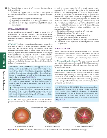
                                                                 rheumatic aortic stenosis there is no commissural fusion
           EFFECTS.  The regurgitant mitral orifice produces     (Fig. 16.32,B).
           progressive increase in left ventricular end-diastolic volume


           Figure 16.32  Aortic valve diseases. Normal aortic valve (A) contrasted with aortic stenosis (B) and aortic insufficiency (C).

