
Page 666 - Textbook of Pathology, 6th Edition
P. 666
650 capillaries is high. Therefore, renal cortex is more prone to
the effects of hypertension.
ii) The renal medulla, on the other hand, is poorly perfused
and any interference in blood supply to it results in medullary
necrosis.
iii) The divisions and subdivisions of the renal artery up to
arterioles are end-arteries and have no anastomoses. Thus,
occlusion of any of the branches results in infarction of the
renal parenchyma supplied by it.
iv) Since the tubular capillary beds are derived from the
efferent arterioles leaving the glomeruli, diseases affecting
the blood flow through glomerular tuft have significant
effects on the tubules as well.
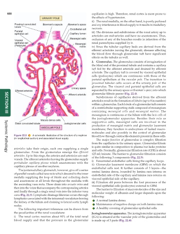
2. Glomerulus. The glomerulus consists of invagination of
the blind end of the proximal tubule and contains a capillary
tuft fed by the afferent arteriole and drained by efferent
arteriole. The capillary tuft is covered by visceral epithelial
cells (podocytes) which are continuous with those of the
parietal epithelium at the vascular pole. The transition to
proximal tubular cells occurs at the urinary pole of the
glomerulus. The visceral and parietal epithelial cells are
separated by the urinary space or Bowman’s space, into which
glomerular filtrate passes (Fig. 22.4).
Subdivisions of capillaries derived from the afferent
arterioles result in the formation of lobules (up to 8 in number)
within a glomerulus. Each lobule of a glomerular tuft consists
of a centrilobular supporting stalk composed of mesangium
containing mesangial cells and mesangial matrix. The
mesangium is continuous at the hilum with the lacis cells of
SECTION III
the juxtaglomerular apparatus. Besides their role as
supportive cells, mesangial cells are involved in the
production of mesangial matrix and glomerular basement
membrane; they function in endocytosis of leaked macro-
molecules and also possibly in the control of glomerular
Figure 22.3 A schematic illustration of the structure of a nephron blood flow through contractile elements present in these cells.
(in longitudinal section) and associated blood supply.
The major function of glomerulus is complex filtration
from the capillaries to the urinary space. Glomerular filtrate
is quite similar in composition to plasma but lacks proteins
arterioles take their origin, each one supplying a single and cells. Normally, glomerular filtration rate (GFR) is about
glomerulus. From the glomerulus emerge the efferent 125 ml/minute. The barrier to glomerular filtration consists
Systemic Pathology
arterioles. Up to this stage, the arteries and arterioles are end- of the following 3 components (Fig. 22.5):
vessels. The efferent arterioles leaving the glomerulus supply i) Fenestrated endothelial cells lining the capillary loops.
peritubular capillary plexus which anastomoses with the ii) Glomerular basement membrane (GBM) on which the
capillary plexus of another nephron. endothelial cells rest. It further consists of 3 layers—the
The juxtamedullary glomeruli, however, give off a series central lamina densa, bounded by lamina rara interna on
of parallel vessels called vasa recta which descend to the inner endothelial side of the capillary and lamina rara externa on
medulla supplying the loop of Henle and collecting ducts visceral epithelial side of the capillary.
and anastomose at all levels throughout the medulla with iii) Filtration slit pores between the foot processes of the
the ascending vasa recta. These drain into arcuate veins and visceral epithelial cells (podocytes) external to GBM.
then into the veins that accompany the corresponding arteries The barrier to filtration of macromolecules of the size and
and finally through a single renal vein into the inferior vena molecular weight of albumin and larger depends upon the
cava (Fig. 22.3). Lymphatic drainage likewise occurs through following:
lymphatics associated with the intrarenal vasculature leaving A normal lamina densa.
the kidney at the hilum and draining to lateral aortic lymph
nodes. Maintenance of negative charge on both lamina rarae.
The following important inferences can be drawn from A healthy covering of glomerular epithelial cells.
the peculiarities of the renal vasculature: Juxtaglomerular apparatus. The juxtaglomerular apparatus
i) The renal cortex receives about 90% of the total renal (JGA) is situated at the vascular pole of the glomerulus and
blood supply and that the pressure in the glomerular is made up of 3 parts (Fig. 22.4):