
Page 667 - Textbook of Pathology, 6th Edition
P. 667
651
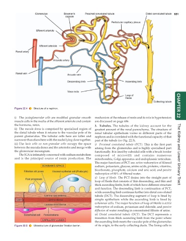
Figure 22.4 Structure of a nephron. CHAPTER 22
i) The juxtaglomerular cells are modified granular smooth mechanism of the release of renin and its role in hypertension
muscle cells in the media of the afferent arteriole and contain are discussed on page 686.
the hormone, renin. 3. Tubules. The tubules of the kidney account for the
ii) The macula densa is comprised by specialised region of greatest amount of the renal parenchyma. The structure of
the distal tubule when it returns to the vascular pole of its renal tubular epithelium varies in different parts of the
parent glomerulus. The tubular cells here are taller and nephron and is correlated with the functional capacity of that
narrower than elsewhere with the nuclei lying close together. part of the tubule (see Fig. 22.3).
iii) The lacis cells or non-granular cells occupy the space i) Proximal convoluted tubule (PCT). This is the first part
between the macula densa and the arterioles and merge with arising from the glomerulus and is highly specialised part
the glomerular mesangium. functionally. It is lined by cuboidal cells with a brush border
The JGA is intimately concerned with sodium metabolism composed of microvilli and contains numerous The Kidney and Lower Urinary Tract
and is the principal source of renin production. The mitochondria, Golgi apparatus and endoplasmic reticulum.
The major functions of PCT are: active reabsorption of filtered
sodium, potassium, glucose, amino acids, proteins, vitamins,
bicarbonate, phosphate, calcium and uric acid, and passive
reabsorption of 80% of filtered water.
ii) Loop of Henle. The PCT drains into the straight part of
loop of Henle that consists of thin descending, and thin and
thick ascending limbs, both of which have different structure
and function. The descending limb is continuation of PCT,
while ascending limb continues further into distal convoluted
tubule (DCT). The descending segment of loop is lined by
simple epithelium while the ascending limb is lined by
columnar cells. The major function of loop of Henle is active
reabsorption of sodium, potassium and chloride, and passive
diffusion of water resulting in concentrated filtrate of urine.
iii) Distal convoluted tubule (DCT). The DCT represents a
transition from thick ascending limb from the point where
the ascending limb meets the vascular pole of the glomerulus
Figure 22.5 Ultrastructure of glomerular filtration barrier. of its origin, to the early collecting ducts. The lining cells in