
Page 663 - Textbook of Pathology, 6th Edition
P. 663
COMPLICATIONS. Late stage of chronic pancreatitis may 647
be complicated by diabetes mellitus, pancreatic insufficiency
with steatorrhoea and malabsorption and formation of
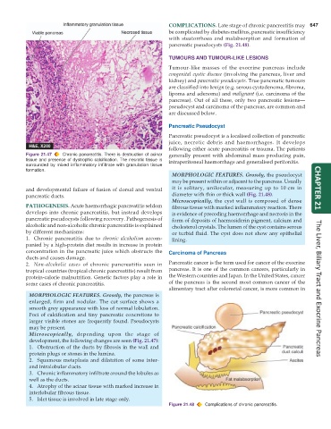
pancreatic pseudocysts (Fig. 21.48).
TUMOURS AND TUMOUR-LIKE LESIONS
Tumour-like masses of the exocrine pancreas include
congenital cystic disease (involving the pancreas, liver and
kidney) and pancreatic pseudocysts. True pancreatic tumours
are classified into benign (e.g. serous cystadenoma, fibroma,
lipoma and adenoma) and malignant (i.e. carcinoma of the
pancreas). Out of all these, only two pancreatic lesions—
pseudocyst and carcinoma of the pancreas, are common and
are discussed below.
Pancreatic Pseudocyst
Pancreatic pseudocyst is a localised collection of pancreatic
juice, necrotic debris and haemorrhages. It develops
following either acute pancreatitis or trauma. The patients
Figure 21.47 Chronic pancreatitis. There is destruction of acinar generally present with abdominal mass producing pain,
tissue and presence of dystrophic calcification. The necrotic tissue is intraperitoneal haemorrhage and generalised peritonitis.
surrounded by mixed inflammatory infiltrate with granulation tissue
formation.
MORPHOLOGIC FEATURES. Grossly, the pseudocyst
may be present within or adjacent to the pancreas. Usually
and developmental failure of fusion of dorsal and ventral it is solitary, unilocular, measuring up to 10 cm in CHAPTER 21
pancreatic ducts. diameter with thin or thick wall (Fig. 21.48).
Microscopically, the cyst wall is composed of dense
PATHOGENESIS. Acute haemorrhagic pancreatitis seldom fibrous tissue with marked inflammatory reaction. There
develops into chronic pancreatitis, but instead develops is evidence of preceding haemorrhage and necrosis in the
pancreatic pseudocysts following recovery. Pathogenesis of form of deposits of haemosiderin pigment, calcium and
alcoholic and non-alcoholic chronic pancreatitis is explained cholesterol crystals. The lumen of the cyst contains serous
by different mechanisms: or turbid fluid. The cyst does not show any epithelial
1. Chronic pancreatitis due to chronic alcoholism accom- lining.
panied by a high-protein diet results in increase in protein
concentration in the pancreatic juice which obstructs the Carcinoma of Pancreas
ducts and causes damage.
2. Non-alcoholic cases of chronic pancreatitis seen in Pancreatic cancer is the term used for cancer of the exocrine
tropical countries (tropical chronic pancreatitis) result from pancreas. It is one of the common cancers, particularly in
protein-calorie malnutrition. Genetic factors play a role in the Western countries and Japan. In the United States, cancer
some cases of chronic pancreatitis. of the pancreas is the second most common cancer of the
alimentary tract after colorectal cancer, is more common in The Liver, Biliary Tract and Exocrine Pancreas
MORPHOLOGIC FEATURES. Grossly, the pancreas is
enlarged, firm and nodular. The cut surface shows a
smooth grey appearance with loss of normal lobulation.
Foci of calcification and tiny pancreatic concretions to
larger visible stones are frequently found. Pseudocysts
may be present.
Microscopically, depending upon the stage of
development, the following changes are seen (Fig. 21.47):
1. Obstruction of the ducts by fibrosis in the wall and
protein plugs or stones in the lumina.
2. Squamous metaplasia and dilatation of some inter-
and intralobular ducts.
3. Chronic inflammatory infiltrate around the lobules as
well as the ducts.
4. Atrophy of the acinar tissue with marked increase in
interlobular fibrous tissue.
5. Islet tissue is involved in late stage only.
Figure 21.48 Complications of chronic pancreatitis.