
Page 665 - Textbook of Pathology, 6th Edition
P. 665
649
The Kidney and
Chapter 22
Chapter 22
Lower Urinary Tract
KIDNEY
NORMAL STRUCTURE
ANATOMY. The kidneys are bean-shaped paired organs,
each weighing about 150 gm in the adult male and about
135 gm in the adult female. The hilum of the kidney is
situated at the midpoint on the medial aspect where the
artery, vein, lymphatics and ureter are located. The kidney
is surrounded by a thin fibrous capsule which is adherent at
the hilum.
Cut surface of the kidney shows 3 main structures: well-
demarcated peripheral cortex, inner medulla and the innermost
renal pelvis (Fig. 22.1):
The renal cortex forms the outer rim of the kidney and is
about 1 cm in thickness. It contains all the glomeruli and CHAPTER 22
about 85% of the nephron tubules. Remaining 15% nephrons
consisting of collecting tubules, collecting ducts, loops of Figure 22.2 Cross-section of the kidney showing arterial blood
Henle and vasa recta send their loops into the medulla, and supply.
are therefore called juxtamedullary nephrons. This latter part
of the cortex forms faint striations called medullary rays, a of each renal pyramid for passage of urine collected from
misnomer since theses structures are located in the cortex collecting ducts and goes down into minor calyces.
but are destined for medulla. Columns of renal cortical tissue The renal pelvis is the funnel-shaped collection area of
that extend into the space between adjacent pyramids are the urine for drainage into the ureter. The minor calyces (8-
called the renal column (septa) of Bertin; they contain the 18 in number in a normal kidney) collect urine from renal
interlobar arteries. papillae and drain into major calyces (2-3 in a normal kidney).
The renal medulla is composed of 8-18 cone-shaped renal HISTOLOGY. The parenchyma of each kidney is composed
pyramids. The base of a renal pyramid lies adjacent to the of approximately one million microstructures called
outer cortex and forms the cortico-medullary junction, while nephrons. A nephron, in turn, consists of 5 major parts, each
the apex of each called the renal papilla contains the opening having a functional role in the formation of urine: the The Kidney and Lower Urinary Tract
glomerular capsule (glomerulus and Bowman’s capsule), the
proximal convoluted tubule (PCT), the loop of Henle, the
distal convoluted tubule (DCT), and the collecting ducts.
From point of view of diseases of the kidneys, 4 components
of renal parenchyma require further elaboration: renal
vasculature, glomeruli, tubules and interstitium.
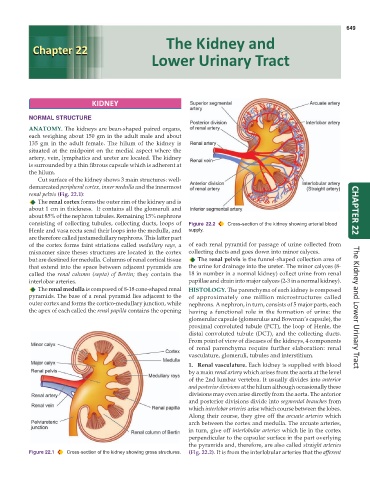
1. Renal vasculature. Each kidney is supplied with blood
by a main renal artery which arises from the aorta at the level
of the 2nd lumbar vertebra. It usually divides into anterior
and posterior divisions at the hilum although occasionally these
divisions may even arise directly from the aorta. The anterior
and posterior divisions divide into segmental branches from
which interlobar arteries arise which course between the lobes.
Along their course, they give off the arcuate arteries which
arch between the cortex and medulla. The arcuate arteries,
in turn, give off interlobular arteries which lie in the cortex
perpendicular to the capsular surface in the part overlying
the pyramids and, therefore, are also called straight arteries
Figure 22.1 Cross-section of the kidney showing gross structures. (Fig. 22.2). It is from the interlobular arteries that the afferent