
Page 668 - Textbook of Pathology, 6th Edition
P. 668
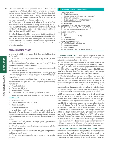
652 DCT are cuboidal. The epithelial cells at the point of TABLE 22.1: Renal Function Tests.
beginning of DCT are taller, narrower and more closely
packed to form the macula densa of JGA as already described. 1. URINE ANALYSIS:
The DCT further contributes to urinary concentration and i) Physical examination
(output, colour, specific gravity, pH, osmolality)
acidification, while the macula densa of JGA is the source of ii) Chemical constituents
renin and has a role in sodium metabolism. (protein, glucose, red cells, haemoglobin)
iv) Collecting ducts. The system of collecting ducts is the final iii) Bacteriologic examination
pathway by which urine reaches the tip of renal papilla. The iv) Microscopy
cells lining the collecting ducts are cuboidal but lack the brush 2. CONCENTRATION AND DILUTION TESTS:
border. Collecting ducts reabsorb water under control of i) Concentration test (fluid deprivation test)
ii)
Dilution test (excess fluid intake test)
+
+
ADH, and secrete H and K ions.
3. BLOOD CHEMISTRY:
4. Interstitium. In health, the renal cortical interstitium is i) Urea
scanty and consists of a small number of fibroblast-like cells. ii) Blood urea nitrogen (BUN)
But the medullary interstitium is more plentiful and contains iii) Creatinine
stellate interstitial cells which are considered to produce an iv) β 2 -microglobulin
anti-hypertensive agent and are involved in the metabolism 4. RENAL CLEARANCE TEST:
i)
Inulin or mannitol clearance test
of prostaglandins. ii) Creatinine clearance
iii) Urea clearance
RENAL FUNCTION TESTS iv) Para-aminohippuric acid (PAH) clearance
In general, the kidney performs the following vital functions
in the body: 1. URINE ANALYSIS. The simplest diagnostic tests for
1. Excretion of waste products resulting from protein renal function is the physical, chemical, bacteriologic and
metabolism. microscopic examination of the urine.
+
2. Regulation of acid-base balance by excretion of H ions i) The physical examination includes 24-hour urinary output,
(acidification) and bicarbonate ions. colour, specific gravity and osmolality. Normally urine is
3. Regulation of salt-water balance by hormones secreted both clear, pale or straw-coloured due to pigment urochrome and
700-2500 ml (average 1200 ml) of urine is passed in 24 hours,
intra- and extra-renally. mostly during day time. Specific gravity is used to measure
SECTION III
4. Formation of renin and erythropoietin and thereby playing the concentrating and diluting power of the kidneys.
a role in the regulation of blood pressure and erythropoiesis ii) The chemical tests are carried out to detect the presence of
respectively. protein, glucose, red cells and haemoglobin to assess the
In order to assess renal function, a number of tests have
been devised which give information regarding the following permeability of glomerular membrane. A number of
convenient dipstick tests are available for testing these
parameters: chemical substances and pH. These consist of paper strips
a) Renal blood flow impregnated with appropriate reagents and indicator dyes.
b) Glomerular filtration
c) Renal tubular function ii) The bacteriologic examination of the urine is done by proper
d) Urinary outflow unhindered by any obstruction. and aseptic collection of midstream specimen of urine.
iv) Urine microscopy is undertaken on a fresh unstained
Renal function tests are broadly divided into 4 groups sample. Various components observed on microscopic
Systemic Pathology
(Table 22.1): examination of the urine in renal disease are red cells, pus
1. Urine analysis. cells, epithelial cells, crystals and urinary casts. The casts are
2. Concentration and dilution tests. moulded into cylindrical shapes by passage along tubules
3. Blood chemistry. in which they are formed. They are the result of precipitation
4. Renal clearance tests. of proteins in the tubule that includes not only albumin but
In addition, renal biopsy is performed to confirm the also the tubular secretion of the Tamm Horsfall protein. The
diagnosis of renal disease. Renal biopsy is ideally fixed in latter is a high molecular weight glycoprotein normally
alcoholic Bouin’s solution and examined by routine morpho- secreted by ascending loop of Henle and DCT and probably
logy combined with special stains and further studies as has body defense function normally. Its secretion is increased
under: in glomerular and tubular diseases. Casts may be hyaline type
1. Periodic acid-Schiff stain for highlighting glomerular consisting of only proteins indicating a non-inflammatory
basement membrane. etiology of glomerular filtration of proteins, leucocyte casts
2. Silver impregnation to outline the glomerular and tubular inflammatory in origin, or red cell casts from haematuria.
basement membrane. 2. CONCENTRATION AND DILUTION TESTS. Concen-
3. Immunofluorescence to localise the antigens, complements tration and dilution tests are designed to evaluate functional
and immunoglobulins. capacity of the renal tubules. The ability of the nephron to
4. Electron microscopy to see the ultrastructure of glomerular concentrate or dilute urine is dependent upon both functional
changes. activity of the tubular cells in the renal medulla and the