
Page 772 - Textbook of Pathology, 6th Edition
P. 772
756
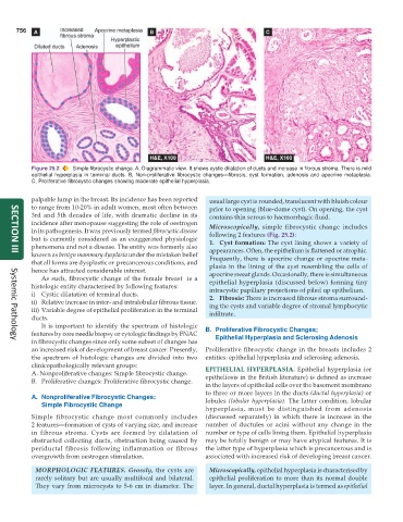
Figure 25.2 Simple fibrocystic change. A, Diagrammatic view. It shows cystic dilatation of ducts and increase in fibrous stroma. There is mild
epithelial hyperplasia in terminal ducts. B, Non-proliferative fibrocystic changes—fibrosis, cyst formation, adenosis and apocrine metaplasia.
C, Proliferative fibrocystic changes showing moderate epithelial hyperplasia.
palpable lump in the breast. Its incidence has been reported usual large cyst is rounded, translucent with bluish colour
to range from 10-20% in adult women, most often between prior to opening (blue-dome cyst). On opening, the cyst
3rd and 5th decades of life, with dramatic decline in its contains thin serous to haemorrhagic fluid.
incidence after menopause suggesting the role of oestrogen Microscopically, simple fibrocystic change includes
in its pathogenesis. It was previously termed fibrocystic disease following 2 features (Fig. 25.2):
but is currently considered as an exaggerated physiologic 1. Cyst formation: The cyst lining shows a variety of
phenomena and not a disease. The entity was formerly also appearances. Often, the epithelium is flattened or atrophic.
known as benign mammary dysplasia under the mistaken belief Frequently, there is apocrine change or apocrine meta-
SECTION III
that all forms are dysplastic or precancerous conditions, and plasia in the lining of the cyst resembling the cells of
hence has attracted considerable interest. apocrine sweat glands. Occasionally, there is simultaneous
As such, fibrocystic change of the female breast is a epithelial hyperplasia (discussed below) forming tiny
histologic entity characterised by following features: intracystic papillary projections of piled up epithelium.
i) Cystic dilatation of terminal ducts. 2. Fibrosis: There is increased fibrous stroma surround-
ii) Relative increase in inter- and intralobular fibrous tissue. ing the cysts and variable degree of stromal lymphocytic
iii) Variable degree of epithelial proliferation in the terminal infiltrate.
ducts.
It is important to identify the spectrum of histologic
features by core needle biopsy or cytologic findings by FNAC B. Proliferative Fibrocystic Changes;
in fibrocystic changes since only some subset of changes has Epithelial Hyperplasia and Sclerosing Adenosis
Systemic Pathology
an increased risk of development of breast cancer. Presently, Proliferative fibrocystic change in the breasts includes 2
the spectrum of histologic changes are divided into two entities: epithelial hyperplasia and sclerosing adenosis.
clinicopathologically relevant groups: EPITHELIAL HYPERPLASIA. Epithelial hyperplasia (or
A. Nonproliferative changes: Simple fibrocystic change. epitheliosis in the British literature) is defined as increase
B. Proliferative changes: Proliferative fibrocystic change.
in the layers of epithelial cells over the basement membrane
to three or more layers in the ducts (ductal hyperplasia) or
A. Nonproliferative Fibrocystic Changes: lobules (lobular hyperplasia). The latter condition, lobular
Simple Fibrocystic Change
hyperplasia, must be distinguished from adenosis
Simple fibrocystic change most commonly includes (discussed separately) in which there is increase in the
2 features—formation of cysts of varying size, and increase number of ductules or acini without any change in the
in fibrous stroma. Cysts are formed by dilatation of number or type of cells lining them. Epithelial hyperplasia
obstructed collecting ducts, obstruction being caused by may be totally benign or may have atypical features. It is
periductal fibrosis following inflammation or fibrous the latter type of hyperplasia which is precancerous and is
overgrowth from oestrogen stimulation. associated with increased risk of developing breast cancer.
MORPHOLOGIC FEATURES. Grossly, the cysts are Microscopically, epithelial hyperplasia is characterised by
rarely solitary but are usually multifocal and bilateral. epithelial proliferation to more than its normal double
They vary from microcysts to 5-6 cm in diameter. The layer. In general, ductal hyperplasia is termed as epithelial