
Page 777 - Textbook of Pathology, 6th Edition
P. 777
761
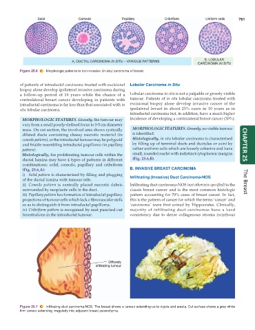
Figure 25.6 Morphologic patterns in non-invasive (in situ) carcinoma of breast.
of patients of intraductal carcinoma treated with excisional Lobular Carcinoma in Situ
biopsy alone develop ipsilateral invasive carcinoma during
a follow-up period of 10 years while the chance of a Lobular carcinoma in situ is not a palpable or grossly visible
contralateral breast cancer developing in patients with tumour. Patients of in situ lobular carcinoma treated with
intraductal carcinoma is far less than that associated with in excisional biopsy alone develop invasive cancer of the
situ lobular carcinoma. ipsilateral breast in about 25% cases in 10 years as in
intraductal carcinoma but, in addition, have a much higher
MORPHOLOGIC FEATURES. Grossly, the tumour may incidence of developing a contralateral breast cancer (30%).
vary from a small poorly-defined focus to 3-5 cm diameter
mass. On cut section, the involved area shows cystically MORPHOLOGIC FEATURES. Grossly, no visible tumour
dilated ducts containing cheesy necrotic material (in is identified.
comedo pattern), or the intraductal tumour may be polypoid Histologically, in situ lobular carcinoma is characterised
and friable resembling intraductal papilloma (in papillary by filling up of terminal ducts and ductules or acini by CHAPTER 25
pattern). rather uniform cells which are loosely cohesive and have
Histologically, the proliferating tumour cells within the small, rounded nuclei with indistinct cytoplasmic margins
ductal lumina may have 4 types of patterns in different (Fig. 25.6,B).
combinations: solid, comedo, papillary and cribriform
(Fig. 25.6,A): B. INVASIVE BREAST CARCINOMA
i) Solid pattern is characterised by filling and plugging
of the ductal lumina with tumour cells. Infiltrating (Invasive) Duct Carcinoma-NOS
ii) Comedo pattern is centrally placed necrotic debris Infiltrating duct carcinoma-NOS (not otherwise specified) is the The Breast
surrounded by neoplastic cells in the duct. classic breast cancer and is the most common histologic
iii) Papillary pattern has formation of intraductal papillary pattern accounting for 70% cases of breast cancer. In fact,
projections of tumour cells which lack a fibrovascular stalk this is the pattern of cancer for which the terms ‘cancer’ and
so as to distinguish it from intraductal papilloma. ‘carcinoma’ were first coined by Hippocrates. Clinically,
iv) Cribriform pattern is recognised by neat punched out majority of infiltrating duct carcinomas have a hard
fenestrations in the intraductal tumour. consistency due to dense collagenous stroma (scirrhous
Figure 25.7 Infiltrating duct carcinoma-NOS. The breast shows a tumour extending up to nipple and areola. Cut surface shows a grey white
firm tumour extending irregularly into adjacent breast parenchyma.