
Page 769 - Textbook of Pathology, 6th Edition
P. 769
753
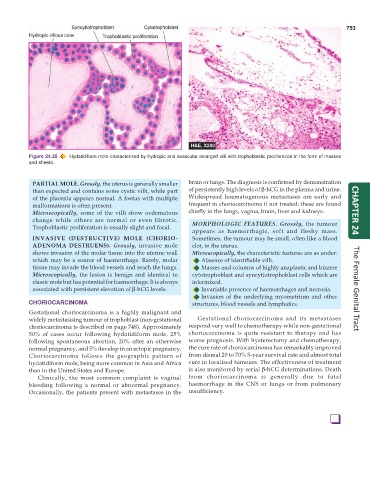
Figure 24.35 Hydatidiform mole characterised by hydropic and avascular enlarged villi with trophoblastic proliferation in the form of masses
and sheets.
PARTIAL MOLE. Grossly, the uterus is generally smaller brain or lungs. The diagnosis is confirmed by demonstration
than expected and contains some cystic villi, while part of persistently high levels of β-hCG in the plasma and urine.
of the placenta appears normal. A foetus with multiple Widespread haematogenous metastases are early and
malformations is often present. frequent in choriocarcinoma if not treated; these are found
Microscopically, some of the villi show oedematous chiefly in the lungs, vagina, brain, liver and kidneys. CHAPTER 24
change while others are normal or even fibrotic. MORPHOLOGIC FEATURES. Grossly, the tumour
Trophoblastic proliferation is usually slight and focal.
appears as haemorrhagic, soft and fleshy mass.
INVASIVE (DESTRUCTIVE) MOLE (CHORIO- Sometimes, the tumour may be small, often like a blood
ADENOMA DESTRUENS). Grossly, invasive mole clot, in the uterus.
shows invasion of the molar tissue into the uterine wall Microscopically, the characteristic features are as under:
which may be a source of haemorrhage. Rarely, molar Absence of identifiable villi.
tissue may invade the blood vessels and reach the lungs. Masses and columns of highly anaplastic and bizarre
Microscopically, the lesion is benign and identical to cytotrophoblast and syncytiotrophoblast cells which are
classic mole but has potential for haemorrhage. It is always intermixed.
associated with persistent elevation of β-hCG levels. Invariable presence of haemorrhages and necrosis. The Female Genital Tract
Invasion of the underlying myometrium and other
CHORIOCARCINOMA structures, blood vessels and lymphatics.
Gestational choriocarcinoma is a highly malignant and
widely metastasising tumour of trophoblast (non-gestational Gestational choriocarcinoma and its metastases
choriocarcinoma is described on page 748). Approximately respond very well to chemotherapy while non-gestational
50% of cases occur following hydatidiform mole, 25% choriocarcinoma is quite resistant to therapy and has
following spontaneous abortion, 20% after an otherwise worse prognosis. With hysterectomy and chemotherapy,
normal pregnancy, and 5% develop in an ectopic pregnancy. the cure rate of choriocarcinoma has remarkably improved
Choriocarcinoma follows the geographic pattern of from dismal 20 to 70% 5-year survival rate and almost total
hydatidiform mole, being more common in Asia and Africa cure in localised tumours. The effectiveness of treatment
than in the United States and Europe. is also monitored by serial β-hCG determinations. Death
Clinically, the most common complaint is vaginal from choriocarcinoma is generally due to fatal
bleeding following a normal or abnormal pregnancy. haemorrhage in the CNS or lungs or from pulmonary
Occasionally, the patients present with metastases in the insufficiency.
❑