
Page 768 - Textbook of Pathology, 6th Edition
P. 768
752
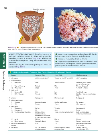
Figure 24.34 Uterus containing hydatidiform mole. The specimen shows numerous, variable-sized, grape-like translucent vesicles containing
clear fluid. Tan areas of haemorrhage are also seen.
COMPLETE (CLASSIC) MOLE. Grossly, the uterus is Large, round, oedematous and acellular villi due to
enlarged and characteristically filled with grape-like hydropic degeneration forming central cisterns.
vesicles up to 3 cm in diameter (Fig. 24.34). The vesicles Decreased vascularity of villous stroma.
contain clear watery fluid. Rarely, a macerated foetus may Trophoblastic proliferation in the form of masses and
be found. sheets of both cytotrophoblast and syncytiotrophoblast,
Microscopically, the features are quite typical. These are generally circumferential around the villi.
as under (Fig. 24.35):
SECTION III
TABLE 24.8: Comparative Features of Major Forms of Gestational Trophoblastic Disease.
Feature Complete Mole Partial Mole Choriocarcinoma
1. Karyotype 46,XX or rarely 46,XY Triploid i.e. 69,XXY or 69,XXX 46,XY or variable
2. Clinical findings
i) Diagnosis Mole Missed abortion Abortion; molar, ectopic
or normal pregnancy
ii) Vaginal bleeding Marked Mild Marked, abnormal
iii) Uterus size Large Small Generally not bulky
3. hCG levels
Systemic Pathology
i) Serum hCG High Low Persistently high
ii) hCG in tissues Marked Mild Localised in syncytiotrophoblast
only
4. Embryo Not present May be present Not present
5. Gross appearance
i) Vesicles Large and regular Smaller and irregular No vesicles
ii) Villi Present Present Always absent
6. Microscopy
i) Villous size Uniform Variable None present
ii) Hydropic villi All Some None
iii) Trophoblastic proliferation Diffuse, all three Focal, syncytiotrophoblast only Both cytotrophoblast
(cytotrophoblast, and syncytiotrophoblast
intermediate trophoblast
and syncytiotrophoblast)
iv) Atypia Diffuse Minimal Marked
v) Blood vessels Generally absent Present Present and abnormal
7. Persistence after initial 20% 7% May metastasise rapidly
therapy if not treated
8. Behaviour 2% may develop Choriocarcinoma almost Survival rate with
choriocarcinoma never develops chemotherapy 70%