
Page 774 - Textbook of Pathology, 6th Edition
P. 774
758
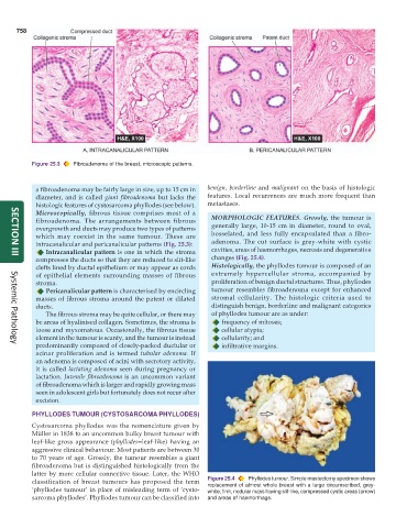
Figure 25.3 Fibroadenoma of the breast, microscopic patterns.
a fibroadenoma may be fairly large in size, up to 15 cm in benign, borderline and malignant on the basis of histologic
diameter, and is called giant fibroadenoma but lacks the features. Local recurrences are much more frequent than
histologic features of cystosarcoma phyllodes (see below). metastases.
Microscopically, fibrous tissue comprises most of a
fibroadenoma. The arrangements between fibrous MORPHOLOGIC FEATURES. Grossly, the tumour is
overgrowth and ducts may produce two types of patterns generally large, 10-15 cm in diameter, round to oval,
which may coexist in the same tumour. These are bosselated, and less fully encapsulated than a fibro-
intracanalicular and pericanalicular patterns (Fig. 25.3): adenoma. The cut surface is grey-white with cystic
Intracanalicular pattern is one in which the stroma cavities, areas of haemorrhages, necrosis and degenerative
compresses the ducts so that they are reduced to slit-like changes (Fig. 25.4).
SECTION III
clefts lined by ductal epithelium or may appear as cords Histologically, the phyllodes tumour is composed of an
of epithelial elements surrounding masses of fibrous extremely hypercellular stroma, accompanied by
stroma. proliferation of benign ductal structures. Thus, phyllodes
Pericanalicular pattern is characterised by encircling tumour resembles fibroadenoma except for enhanced
masses of fibrous stroma around the patent or dilated stromal cellularity. The histologic criteria used to
ducts. distinguish benign, borderline and malignant categories
The fibrous stroma may be quite cellular, or there may of phyllodes tumour are as under:
be areas of hyalinised collagen. Sometimes, the stroma is frequency of mitoses;
loose and myxomatous. Occasionally, the fibrous tissue cellular atypia;
element in the tumour is scanty, and the tumour is instead cellularity; and
predominantly composed of closely-packed ductular or infiltrative margins.
Systemic Pathology
acinar proliferation and is termed tubular adenoma. If
an adenoma is composed of acini with secretory activity,
it is called lactating adenoma seen during pregnancy or
lactation. Juvenile fibroadenoma is an uncommon variant
of fibroadenoma which is larger and rapidly growing mass
seen in adolescent girls but fortunately does not recur after
excision.
PHYLLODES TUMOUR (CYSTOSARCOMA PHYLLODES)
Cystosarcoma phyllodes was the nomenclature given by
Müller in 1838 to an uncommon bulky breast tumour with
leaf-like gross appearance (phyllodes=leaf-like) having an
aggressive clinical behaviour. Most patients are between 30
to 70 years of age. Grossly, the tumour resembles a giant
fibroadenoma but is distinguished histologically from the
latter by more cellular connective tissue. Later, the WHO
classification of breast tumours has proposed the term Figure 25.4 Phyllodes tumour. Simple mastectomy specimen shows
replacement of almost whole breast with a large circumscribed, grey-
‘phyllodes tumour’ in place of misleading term of ‘cysto- white, firm, nodular mass having slit-like, compressed cystic areas (arrow)
sarcoma phyllodes’. Phyllodes tumour can be classified into and areas of haemorrhage.