
Page 821 - Textbook of Pathology, 6th Edition
P. 821
805
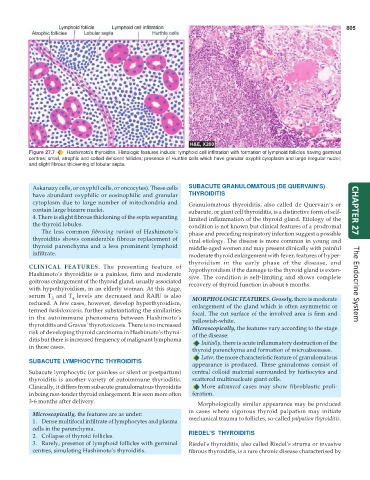
Figure 27.7 Hashimoto’s thyroiditis. Histologic features include: lymphoid cell infiltration with formation of lymphoid follicles having germinal
centres; small, atrophic and colloid-deficient follicles; presence of Hurthle cells which have granular oxyphil cytoplasm and large irregular nuclei;
and slight fibrous thickening of lobular septa.
Askanazy cells, or oxyphil cells, or oncocytes). These cells SUBACUTE GRANULOMATOUS (DE QUERVAIN’S)
have abundant oxyphilic or eosinophilic and granular THYROIDITIS
cytoplasm due to large number of mitochondria and Granulomatous thyroiditis, also called de Quervain’s or
contain large bizarre nuclei. subacute, or giant cell thyroiditis, is a distinctive form of self- CHAPTER 27
4. There is slight fibrous thickening of the septa separating limited inflammation of the thyroid gland. Etiology of the
the thyroid lobules. condition is not known but clinical features of a prodromal
The less common fibrosing variant of Hashimoto’s phase and preceding respiratory infection suggest a possible
thyroiditis shows considerable fibrous replacement of viral etiology. The disease is more common in young and
thyroid parenchyma and a less prominent lymphoid middle-aged women and may present clinically with painful
infiltrate. moderate thyroid enlargement with fever, features of hyper-
thyroidism in the early phase of the disease, and
CLINICAL FEATURES. The presenting feature of hypothyroidism if the damage to the thyroid gland is exten-
Hashimoto’s thyroiditis is a painless, firm and moderate sive. The condition is self-limiting and shows complete
goitrous enlargement of the thyroid gland, usually associated recovery of thyroid function in about 6 months.
with hypothyroidism, in an elderly woman. At this stage, The Endocrine System
serum T and T levels are decreased and RAIU is also MORPHOLOGIC FEATURES. Grossly, there is moderate
3
4
reduced. A few cases, however, develop hyperthyroidism, enlargement of the gland which is often asymmetric or
termed hashitoxicosis, further substantiating the similarities focal. The cut surface of the involved area is firm and
in the autoimmune phenomena between Hashimoto’s yellowish-white.
thyroiditis and Graves’ thyrotoxicosis. There is no increased Microscopically, the features vary according to the stage
risk of developing thyroid carcinoma in Hashimoto’s thyroi- of the disease:
ditis but there is increased frequency of malignant lymphoma Initially, there is acute inflammatory destruction of the
in these cases.
thyroid parenchyma and formation of microabscesses.
Later, the more characteristic feature of granulomatous
SUBACUTE LYMPHOCYTIC THYROIDITIS
appearance is produced. These granulomas consist of
Subacute lymphocytic (or painless or silent or postpartum) central colloid material surrounded by histiocytes and
thyroiditis is another variety of autoimmune thyrioditis. scattered multinucleate giant cells.
Clinically, it differs from subacute granulomatous thyroiditis More advanced cases may show fibroblastic proli-
in being non-tender thyroid enlargement. It is seen more often feration.
3-6 months after delivery.
Morphologically similar appearance may be produced
Microscopically, the features are as under: in cases where vigorous thyroid palpation may initiate
1. Dense multifocal infiltrate of lymphocytes and plasma mechanical trauma to follicles, so-called palpation thyroiditis.
cells in the parenchyma.
2. Collapse of thyroid follicles. RIEDEL’S THYROIDITIS
3. Rarely, presence of lymphoid follicles with germinal Riedel’s thyroiditis, also called Riedel’s struma or invasive
centres, simulating Hashimoto’s thyroiditis. fibrous thyroiditis, is a rare chronic disease characterised by