
Page 822 - Textbook of Pathology, 6th Edition
P. 822
806 stony-hard thyroid that is densely adherent to the adjacent i) Thyroid-stimulating immunoglobulin (TSI): It binds to TSH
structures in the neck. The condition is clinically significant receptor and stimulates increased release of thyroid hormone.
due to compressive clinical features (e.g. dysphagia, ii) Thyroid growth-stimulating immunoglobulins (TGI): It
dyspnoea, recurrent laryngeal nerve paralysis and stridor) stimulates proliferation of follicular epithelium.
and resemblance with thyroid cancer. Riedel’s struma is seen iii) TSH-binding inhibitor immunoglobulins (TBII): It is
more commonly in females in 4th to 7th decades of life. The inhibitory to binding of TSH to its own receptor. Depending
etiology is unknown but possibly Riedel’s thyroiditis is a part upon its action as inhibitory or stimulatory to follicular
of multifocal idiopathic fibrosclerosis (page 591). This group of epithelium, it may result in alternate episodes of hypo- and
disorders includes: idiopathic retroperitoneal, mediastinal hyperthyroidism.
and retro-orbital fibrosis, and sclerosing cholangitis, all of However, it is not quite clear what stimulates B cells to
which may occur simultaneously with Riedel’s thyroiditis. form these autoantibodies in Graves’ disease. Possibly,
intrathyroidal CD4+ helper T cells are responsible for
MORPHOLOGIC FEATURES. Grossly, the thyroid gland stimulating B cells to secrete autoantibodies.
is usually contracted, stony-hard, asymmetric and firmly
adherent to the adjacent structures. Cut section is hard The pathogenesis of Graves’ infiltrative ophthalmopathy
and devoid of lobulations. is also of autoimmune origin. The evidence in support is the
Microscopically, there is extensive fibrocollagenous intense lymphocytic infiltrate around the ocular muscles and
replacement, marked atrophy of the thyroid parenchyma, detection of circulating autoantibodies against muscle
focally scattered lymphocytic infiltration and invasion of antigen that cross-react with thyroid microsomes.
the adjacent muscle tissue by the process. MORPHOLOGIC FEATURES. Grossly, the thyroid is
moderately, diffusely and symmetrically enlarged and
GRAVES’ DISEASE (DIFFUSE TOXIC GOITRE) may weigh up to 70-90 gm. On cut section, the thyroid
Graves’ disease, also known as Basedow’s disease, primary parenchyma is typically homogeneous, red-brown and
hyperplasia, exophthalmic goitre, and diffuse toxic goitre, is meaty and lacks the normal translucency.
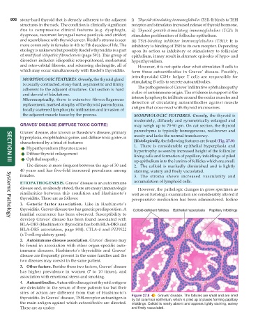
characterised by a triad of features: Histologically, the following features are found (Fig. 27.8):
Hyperthyroidism (thyrotoxicosis) 1. There is considerable epithelial hyperplasia and
Diffuse thyroid enlargement hypertrophy as seen by increased height of the follicular
lining cells and formation of papillary infoldings of piled
Ophthalmopathy.
up epithelium into the lumina of follicles which are small.
SECTION III
The disease is more frequent between the age of 30 and 2. The colloid is markedly diminished and is lightly
40 years and has five-fold increased prevalence among staining, watery and finely vacuolated.
females. 3. The stroma shows increased vascularity and
ETIOPATHOGENESIS. Graves’ disease is an autoimmune accumulation of lymphoid cells.
disease and, as already stated, there are many immunologic However, the pathologic changes in gross specimen as
similarities between this condition and Hashimoto’s well as on histologic examination are considerably altered if
thyroiditis. These are as follows: preoperative medication has been administered. Iodine
1. Genetic factor association. Like in Hashimoto’s
thyroiditis. Graves’disease too has genetic predisposition. A
familial occurrence has been observed. Susceptibility to
develop Graves’ disease has been found associated with
Systemic Pathology
HLA-DR3 (Hashimoto’s thyroiditis has both HLA-DR3 and
HLA-DR5 association, page 804), CTLA-4 and PTPN22
(a T-cell regulatory gene).
2. Autoimmune disease association. Graves’ disease may
be found in association with other organ-specific auto-
immune diseases. Hashimoto’s thyroiditis and Graves’
disease are frequently present in the same families and the
two diseases may coexist in the same patient.
3. Other factors. Besides these two factors, Graves’ disease
has higher prevalence in women (7 to 10 times), and
association with emotional stress and smoking.
4. Autoantibodies. Autoantibodies against thyroid antigens
are detectable in the serum of these patients too but their
sites of action are different from that of Hashimoto’s
thyroiditis. In Graves’ disease, TSH-receptor autoantigen is Figure 27.8 Graves’ disease. The follicles are small and are lined
by tall columnar epithelium, which is piled up at places forming papillary
the main antigen against which autoantibodies are directed. infoldings. Colloid is nearly absent and appears lightly staining, watery
These are as under: and finely vacuolated.