


Page 89 - Textbook of Pathology, 6th Edition
P. 89
myopathy, pericardial effusion in advanced disease as a window period, ELISA may be false positive in autoanti- 73
reaction to opportunistic infection, lymphoma and Kaposi’s bodies, liver disease, recent vaccination against flu, and other
sarcoma. viral infections.
12. Ophthalmic lesions. HIV associated ocular manifes- b) Western blot. If ELISA is positive, confirmation is done by
tations occur from opportunistic infections (e.g. CMV Western blot for presence of specific antibodies against all
retinitis), HIV retinopathy, and secondary tumours. three HIV antigens: gag, pol and env. CHAPTER 4
13. Musculoskeletal lesions. These include osteoporosis, ii) Direct detection of HIV: These tests are as follows:
osteopaenia, septic arthritis, osteomyelitis and polymyositis. a) p24 antigen capture assay.
14. Endocrine lesions. Several metabolic derangements may b) HIV RNA assay methods by reverse transcriptase (RT)
occur during the course of disease. There is syndrome of PCR, branched DNA, nucleic acid sequence-based
lipodystrophy (buffalo hump) due to dyslipidaemia, amplification (NucliSens).
hyperinsulinaemia and hyperglycaemia. There may be c) DNA-PCR by amplification of proviral DNA.
abnormality of thyroid function, hypogonadism and d) Culture of HIV from blood monocytes and CD4+ T cells.
inappropriate release of ADH.
2. Tests for defects in immunity: These tests are used for
LESIONS AND MANIFESTATIONS IN PAEDIATRIC diagnosis as well as for monitoring treatment of cases.
AIDS. Children develop clinical manifestations of AIDS more i) CD4+ T cell counts. Progressive fall in number of CD4+ T
rapidly than adults. Besides development of opportunistic cells is of paramount importance in diagnosis and staging as
infections and tumours, neurologic impairment in children CDC categories described above.
causes slowing of development and growth. ii) Rise in CD8+ T cells.
DIAGNOSIS OF HIV/AIDS. The investigations of a iii) Reversal of CD4+ to CD8+ T cell ratio. Immunopathology Including Amyloidosis
suspected case of HIV/AIDS are categorised into 3 groups: iv) Lymphopenia.
tests for establishing HIV infection, tests for defects in v) Polyclonal hypergammaglobulinaemia.
immunity, and tests for detection of opportunistic infections vi) Increased β-2 microglobulin levels.
and secondary tumours. However, usually initial testing for vii)Platelet count revealing thrombocytopenia.
antibodies is done against HIV by ELISA and confirmation 3. Tests for detection of opportunistic infections and
by Western blot or immunofluorescence test. These tests are secondary tumours: Diagnosis of organs involved in
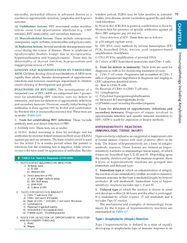
as under (Table 4.6): opportunistic infection and specific tumours secondary to
1. Tests for establishing HIV infection: These include HIV/AIDS is made by aspiration or biopsy methods.
antibody tests and direct detection of HIV.
i) Antibody tests: These tests are as under: HYPERSENSITIVITY REACTIONS
a) ELISA. Initial screening is done by serologic test for (IMMUNOLOGIC TISSUE INJURY)
antibodies by enzyme-linked immunosorbent assay (ELISA) Hypersensitivity is defined as an exaggerated or inappropriate state
against gag and env proteins. The term window period is used of normal immune response with onset of adverse effects on the
for the initial 2 to 4 weeks period when the patient is body. The lesions of hypersensitivity are a form of antigen-
infectious but the screening test is negative, while serocon- antibody reaction. These lesions are termed as hyper-
version is the term used for appearance of antibodies. Besides sensitivity reactions or immunologic tissue injury, of which
4 types are described: type I, II, III and IV. Depending upon
TABLE 4.6: Tests for Diagnosis of HIV/AIDS. the rapidity, duration and type of the immune response, these
1. TESTS FOR ESTABLISHING HIV INFECTION: 4 types of hypersensitivity reactions are grouped into
i) Antibody tests: immediate and delayed type:
a) ELISA 1. Immediate type in which on administration of antigen,
b) Western blot the reaction occurs immediately (within seconds to minutes).
ii) Direct detection of HIV
a) p24 antigen capture assay Immune response in this type is mediated largely by humoral
b) HIV RNA assay antibodies (B cell mediated). Immediate type of hyper-
c) DNA-PCR sensitivity reactions includes type I, II and III.
d) Culture of HIV
2. Delayed type in which the reaction is slower in onset
2. TESTS FOR DEFECTS IN IMMUNITY: and develops within 24-48 hours and the effect is prolonged.
i) CD4+ T cell count: Fall
ii) CD8+ cell count: Increased It is mediated by cellular response (T cell mediated) and it
iii) Ratio of CD4+ T cell/CD8+ T cell count: Reversed includes Type IV reaction.
iv) Lymphopenia The mechanisms and examples of immunologic tissue
v) Hypergammaglobulinaemia injury by the 4 types of hypersensitivity reactions are
vi) Increased β-2 microglobulin level
vii) Platelet count: Thrombocytopenia summarised in Table 4.7.
3. TESTS FOR DETECTION OF OPPORTUNISTIC INFECTION Type I: Anaphylactic (Atopic) Reaction
AND SECONDARY TUMOURS:
i) FNAC Type I hypersensitivity is defined as a state of rapidly
ii) Biopsy
developing or anaphylactic type of immune response to an

