


Page 84 - Textbook of Pathology, 6th Edition
P. 84
68 together comprise about 50% of all HIV positive cases (mostly  These genes and viral components act as markers for the
           contracted sexually), while North-East state of Manipur  laboratory diagnosis of HIV infection. Besides, there is tat
           accounts for 8% of all cases (mostly among intravenous drug  (transcription activator) gene for viral functions such as
           abusers).                                           amplification of viral genes, viral budding and replication.
           ETIOLOGIC AGENT. AIDS is caused by an RNA retrovirus  ROUTES OF TRANSMISSION.  Transmission of HIV
           called human immunodeficiency virus (HIV) which is a type  infection occurs by one of following three routes:
           of human T cell leukaemia-lymphoma virus (HTLV).    1. Sexual transmission. Sexual contact in the main mode
              HIV resembles other HTLVs in shape and size and both  of spread and constitutes 75% of all cases of HIV
           have tropism for CD4 molecules present on subpopulation of  transmission. Most cases of AIDS in the industrialised world
     SECTION I
           T cells which are the particular targets of attack by HIV.  like in the US occur in  homosexual or bisexual males while
           However, HIV differs from HTLV in being cytolytic for T cells  heterosexual promiscuity seems to be the dominant mode of
           causing immunodeficiency (cytopathic virus) while HTLV may  HIV infection in Africa and Asia. Other sexually transmitted
           transform the target cells into T cell leukaemia (transforming virus)  diseases (STDs) may act as cofactors for spread of HIV, in
           (Chapter 8). Two forms of HIV have been described, HIV1  particular gonorrhoeal and chlamydial infection.
           being the etiologic agent for AIDS in the US and Central  Transmission from male-to-male and male-to-female is more
           Africa, while HIV2 causes a similar disease in West Africa  potent route than that from female-to-male.
           and parts of India. Both HIV1 and HIV2 are zoonotic
           infections and their origin can be traced to a species of  2. Transmission via blood and blood products. This mode
           chimpanzees who are natural reservoir of HIV and most  of transmission is the next largest group (25%) and occurs in
           likely source of original infection.                3 groups of high-risk populations:
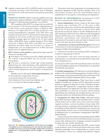
           HIV-I virion or virus particle is spherical in shape and 100-  i) Intravenous drug abusers by sharing needles, syringes etc
           140 nm in size (Fig. 4.4):                          comprise a large group in the US.
              It contains a core having core proteins, chiefly p24 and p18,  ii) Haemophiliacs who have received large amounts of
           two strands of genomic RNA and the enzyme, reverse  clotting factor concentrates from pooled blood components
           transcriptase.                                      from multiple donors.
              The core is covered by a double layer of lipid membrane  iii) Recipients of HIV-infected blood and blood products who have
           derived from the outer membrane of the infected host cell  received multiple transfusions of whole blood or components
           during budding process of virus. The membrane is studded  like platelets and plasma.
           with 2 envelope glycoproteins, gp120 and gp41, in the positions
     General Pathology and Basic Techniques
           shown.                                              3. Perinatal transmission. HIV infection occurs from infected
              Besides various other genes, three important genes code  mother to the newborn during pregnancy transplacentally, or
           for the respective components of virion:            in immediate post-partum period through contamination with
           i) gag (group antigen) for core proteins,           maternal blood, infected amniotic fluid or breast milk.
           ii) pol (polymerase) for reverse transcriptase, and  4. Occupational transmission. There have been a small
           iii) env (envelope) for the envelope proteins.      number of health care workers (HCW), laboratory workers
                                                               and those engaged in disposal of waste of sharps who have
                                                               developed HIV infection by occupational exposure to HIV-
                                                               infected material. It is imperative that these workers follow
                                                               CDC guidelines for  universal precautions which include
                                                               disinfecting and sterilizing all reusable devices and use of
                                                               bleaching solution for disinfecting all blood spillage.
                                                               5. Transmission by other body fluids. Although besides
                                                               blood, HIV has been isolated and identified from a number
                                                               of body fluids such as saliva, tears, sweat and urine, semen,
                                                               vaginal secretions, cervical secretions, breast milk, CSF,
                                                               synovial, pleural, peritoneal and pericardial fluid, there is
                                                               no definite evidence that HIV transmission can occur by any
                                                               of these fluids; isolated cases of such infection reported are
                                                               in likelihood due to concomitant contamination with HIV-
                                                               infected blood.
                                                                  It may, however, be understood regarding spread of HIV
                                                               infection that AIDS cannot be transmitted by casual non-sexual
                                                               contact like shaking hands, hugging, sharing household
                                                               facilities like beds, toilets, utensils etc.
           Figure 4.4  Schematic representation of HIV virion or virus particle.  It should also be appreciated that HIV contaminated
           The particle has core containing proteins, p24 and p18, two strands of  waste products can be sterilised and disinfected by most of the
           viral RNA, and enzyme reverse transcriptase. Bilayer lipid membrane is  chemical germicides used in laboratories at a much lower
           studded with 2 viral glycoproteins, gp120 and gp41, in the positions  concentration. These are: sodium hypochlorite (liquid
           shown.

