


Page 85 - Textbook of Pathology, 6th Edition
P. 85
chlorine bleach), formaldehyde (5%), ethanol (70%),                                                      69
           glutaraldehyde (2%), β-propionolactone. HIV is also heat-
           sensitive and can be inactivated at 56°C for 30 min.
           PATHOGENESIS.  The pathogenesis of HIV infection is
           largely related to the depletion of CD4+ T cells (helper T cells)
           resulting in profound immunosuppression.                                                                   CHAPTER 4
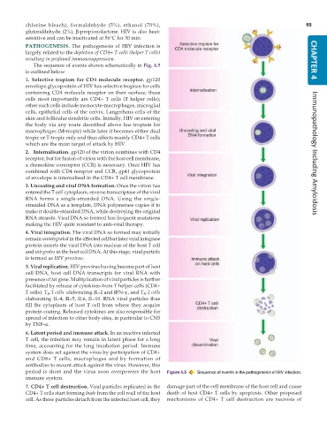
              The sequence of events shown schematically in Fig. 4.5
           is outlined below:
           1. Selective tropism for CD4 molecule receptor. gp120
           envelope glycoprotein of HIV has selective tropism for cells
           containing CD4 molecule receptor on their surface; these
           cells most importantly are CD4+ T cells (T helper cells);
           other such cells include monocyte-macrophages, microglial
           cells, epithelial cells of the cervix, Langerhans cells of the
           skin and follicular dendritic cells. Initially, HIV on entering
           the body via any route described above has tropism for
           macrophages (M-tropic) while later it becomes either dual
           tropic or T-tropic only and thus affects mainly CD4+ T cells
           which are the main target of attack by HIV.
           2. Internalisation. gp120 of the virion combines with CD4                                                  Immunopathology Including Amyloidosis
           receptor, but for fusion of virion with the host cell membrane,
           a chemokine coreceptor (CCR) is necessary. Once HIV has
           combined with CD4 receptor and CCR, gp41 glycoprotein
           of envelope is internalised in the CD4+ T cell membrane.
           3. Uncoating and viral DNA formation. Once the virion has
           entered the T cell cytoplasm, reverse transcriptase of the viral
           RNA forms a single-stranded DNA. Using the single-
           stranded DNA as a template, DNA polymerase copies it to
           make it double-stranded DNA, while destroying the original
           RNA strands. Viral DNA so formed has frequent mutations
           making the HIV quite resistant to anti-viral therapy.
           4. Viral integration. The viral DNA so formed may initially
           remain unintegrated in the affected cell but later viral integrase
           protein inserts the viral DNA into nucleus of the host T cell
           and integrates in the host cell DNA. At this stage, viral particle
           is termed as HIV provirus.
           5. Viral replication. HIV provirus having become part of host
           cell DNA, host cell DNA transcripts for viral RNA with
           presence of tat gene. Multiplication of viral particles is further
           facilitated by release of cytokines from T helper cells (CD4+
           T cells): T  1 cells  elaborating IL-2 and IFN-γ., and T  2 cells
                   H
                                                       H
           elaborating IL-4, IL-5, IL6, IL-10. RNA viral particles thus
           fill the cytoplasm of host T cell from where they acquire
           protein coating. Released cytokines are also responsible for
           spread of infection to other body sites, in particular to CNS
           by TNF-α.
           6. Latent period and immune attack. In an inactive infected
           T cell, the infection may remain in latent phase for a long
           time, accounting for the long incubation period. Immune
           system does act against the virus by participation of CD4+
           and CD8+ T cells, macrophages and by formation of
           antibodies to mount attack against the virus. However, this
           period is short and the virus soon overpowers the host  Figure 4.5  Sequence of events in the pathogenesis of HIV infection.
           immune system.
           7. CD4+ T cell destruction. Viral particles replicated in the  damage part of the cell membrane of the host cell and cause
           CD4+ T cells start forming buds from the cell wall of the host  death of host CD4+ T cells by apoptosis. Other proposed
           cell. As these particles detach from the infected host cell, they  mechanisms of CD4+ T cell destruction are necrosis of

