
Page 5 - Human Environment Interface (3)
P. 5
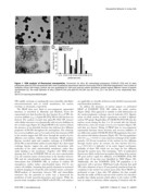
Nanoparticle Behavior in Living Cells

Figure 1. TEM analysis of fluorescent nanoparticles. Fluorescent (A) silica, (B) carboxylated polystyrene (COOH-PS [YO]) and (C) plain
polystyrene (plain-PS [YG]) nanoparticles (NPs) were visualized by transmission electron microscopy (TEM) at 250K-fold magnification. From a series of
randomly chosen TEM images, particle size was quantitated (n.200 each) and the relative abundance plotted against different classes of particle
size/diameter (D). The mean diameter of silica, COOH-PS [YO] and plain-PS [YG] NPs was 4667 nm, 2267 nm and 4565 nm, respectively. Bars,
50 nm.
doi:10.1371/journal.pone.0062018.g001

NPs rapidly exchange at topologically more immobile subcellular        are applicable on virtually all fluorescently labelled nanomaterials
microenvironments such as vesicle membranes, the nuclear               and biomedical problems.
envelope or chromatin, respectively.
                                                                          To address the dynamics of nuclear import we performed
   The FRAP data were fitted to two-component exponential              FRAP of COOH-PS [YO] NPs within the entire nucleus
functions as described in Materials and Methods. From the              (Figure 3D,E). This approach is suitable to yield information on
exponential term that describes the slow fraction (Fslow) of NPs, the  the nucleocytoplasmic exchange rates of molecules [31]. Quanti-
recovery halftime (t50%) of plain-PS [YG] NPs for this fraction was    tation of whole nucleus bleach experiments revealed a biphasic
derived. The analysis revealed that plain-PS [YG] NPs interact         nuclear NP uptake. Rapid influx of COOH-PS [YO] NPs into the
with cellular structures very dynamically with recovery halftimes in   nucleus occurs during the first 5 to 10 seconds after the bleach,
the seconds range (Figure 3C). Rapid FRAP in the nucleus without       followed by a slow, but steady recovery over minutes (Figures 3D,
the presence of an immobile fraction also indicates free diffusion     and data not shown). Fitting of FRAP data to two-component
properties of PS-NPs throughout the nucleoplasm. The relatively        exponential functions shows fractions and recovery halftimes of
low recovery-halftime (one to 2 seconds) of the slow fraction of NPs   two differently mobile COOH-PS [YO] NP populations that enter
in the nucleus suggest that NPs move through the nucleus by free       the nucleus with distinct dynamics (Figure 3E). The fast population
diffusion and random collision with chromatin. FRAP analyses           (1665%, t50% = 3.060.5 s) may represent exchange of the smaller
likewise revealed rapid and complete exchange of COOH-PS               particles (10–20 nm) in-and-out of the nucleus by free diffusion
[YO] NPs at mitochondria within seconds (Figure 3C). Such rapid        through nuclear pores. In contrast, the slow fraction (6365%,
exchange kinetics suggest fast and transient interactions with         t50% = 251642 s) is indicative of an active import mechanism,
molecules of the outer mitochondrial membrane, rather than             probably of those NPs which are too large for free diffusion
stable binding to the membrane. Together with TEM results              through the nuclear pore. The remaining ,20% of unbleached
showing that 50 nm COOH-PS NPs do not enter mitochondria               fluorescence likely results from incomplete bleaching of the nuclear
[17], the transient nature of interactions at the interface between    NP pool and/or rapid free diffusion of COOH-PS [YO] NPs
NPs and the mitochondrial membrane might explain previous              smaller than 10 nm that were identified by TEM (Figure 1D).
observations that COOH-PS NPs, in contrast to cationic PS              Additionally, the fast COOH-PS [YO] NP population is in
nanospheres, neither induce mitochondrial injury [16,17], nor          agreement with a previous TEM-based study showing that gold
reduce cell viability. Assuming that particle dynamics as measured     NPs with a diameter of up to 39 nm passively translocate from the
by kinetic imaging are of specific and consistent nature, systematic   cytoplasm to the nucleus via nuclear pores [32]. Since FRAP was
FRAP-analysis of interactions between NPs and intracellular            performed at least 1 hour after NP addition, the data actually
structures has the potential to evolve into a framework for            describe the traffic of NPs in and out of the nucleus. It is therefore
prediction of both nano-bio-interaction and toxicity. While the        possible that cellular proteins which adhere to the surface of NPs
present study exemplary shows FRAP with PS NPs, such analyses          may be subject to aberrant cellular targeting. Consistent with the

PLOS ONE | www.plosone.org                                             3 April 2013 | Volume 8 | Issue 4 | e62018