
Page 6 - Human Environment Interface (3)
P. 6
Nanoparticle Behavior in Living Cells
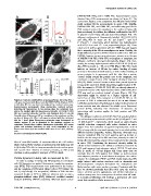
Figure 2. Cellular distribution of nanoparticles. (A) Living HEp-2 (DMEM/10% FBS), and in 100% FBS. Autocorrelation curves
cells were incubated with fluorescent (A) COOH-PS [YO] , (C) plain-PS YG obtained from FCS measurements are shown in Figure 4C. The
or (D) silica NPs for one hour and analyzed by confocal microscopy. curves were fitted to a one-component-free-diffusion model which
Representative micrographs show mid-nuclear confocal sections yields excellent fits for measurements in water, PBS, DMEM,
detecting NP fluorescence in the nucleus (Nu), the cytoplasm (Cy), DMEM/10% FBS, and 100% FBS, and sufficiently good fits for
and in the medium (Me). (B) Fluorescence intensity of COOH-PS [YO] FCS measurements in the cytoplasm or the nucleus of living cells
NPs was recorded along the red line and displayed. Bars, 10 c¸m. (E) (data not shown). In addition, the diffusion coefficient for free GFP
Quantification of fluorescent NP-accumulation in the cytoplasm and the in solution and in living cells was determined (Figure 4D). The
nucleus of living cells. HEp-2 cells were incubated with the indicated diffusion coefficients of fluorescently labelled COOH-PS [YO]
NPs for 1 hour. Mean fluorescence intensities were determined in and silica NPs in water are 8.1 (61.1) mm2s21 and 12.4
circular regions (2 c¸m in diameter) in the medium, nucleus or (62.3) mm2s21, respectively, which translate into hydrodynamic
cytoplasm. Graphs show relative fluorescence intensities (RFI) mean radii of 2964 nm and 1764 nm, respectively (Figure 4E). These
values and standard deviations (n = cells each) normalized to the values are in perfect agreement with the TEM data and suggest
fluorescence intensity in the medium which was set to 2. A.U., arbitrary that a majority of the NPs is monodisperse in water. Notably, the
units. in vitro diffusion properties of NPs remained unaffected in PBS and
doi:10.1371/journal.pone.0062018.g002 DMEM, however, as soon as proteins were present, as in the case
of DMEM/10% FBS, 100% FBS, nucleoplasm or cytoplasm, the
idea of controlled transfer of nanomaterials to the cell nucleus diffusion coefficient decreased dramatically (Figure 4D). Con-
whole nucleus FRAP analyses as performed in this study may aid versely, the average hydrodynamic radius of COOH-PS [YO] and
to (i) develop NPs able to carry macromolecules via nuclear pores silica NPs increased to ,90 nm in FBS (Figure 4E). The results
and execute their delivery to nuclear processes, as well as (ii) indicate an increase of NP-size by surface binding of serum
elucidate nucleocytoplasmic transport of NPs in depth. proteins or formation of small clusters, or both. Surface binding of
serum proteins is in agreement with the idea that a protein
Particle dynamics in living cells as measured by FCS ’corona’ builds around the particle core in the cytoplasm, and
In order to analyse in living cells NP-properties in correlation constitutes a major element of the biological identity of respective
NPs [13]. While ’corona’ formation slows down particle dynamics
with NP-dynamics we applied fluorescence correlation spectros- it may at the same time inhibit intracellular agglomeration of PS-
copy (FCS). FCS measurements were performed in the nucleus NPs. In contrast to COOH-PS [YO] NPs, we already observed a
and the cytoplasm of NP-loaded HEp-2 cells at 25uC (Figure 4A substantial increase of the hydrodynamic radius of silica NPs when
and B, respectively). For comparison, diffusion of NPs was also PBS was used as a solvent instead of water (Figure 4E). This
measured in vitro at 25uC in different solutions such as water, observation might be related to a higher capacity of self-
phosphate-buffered saline (PBS), Dulbecco’s modified essential interaction of silica NPs compared to COOH-PS NPs in the
medium (DMEM), DMEM containing 10% fetal bovine serum presence of PBS. A substantial decrease of the in vitro diffusion
coefficient and increase of hydrodynamic radius in the presence of
serum proteins was also observed for soluble green fluorescent
protein (GFP), indicating that obstruction of dynamics by
interactions with proteins applies for both proteins and NPs
(Figure 4D).
The diffusion coefficient of COOH-PS [YO] and silica-NPs in
the cell nucleus as determined by FCS was 0.8 (60.3) mm2s21 and
1.2 (60.3) mm2s21, respectively, which equals an approximately
ten-fold reduction compared with values obtained in water
(Figure 4D). For evaluation of apparent hydrodynamic radii of
NPs in living cells, the increased viscosity due to immobile cellular
diffusion barriers (organelles, vesicles, filaments, chromatin etc.)
must be considered [33]. For inert particles, such as dextran or
ficoll in the size range of the NPs used here, viscosity in nuclei of
living cells is increased by a factor of four compared to the viscosity
in water [34]. Considering the increased viscosity within the
cellular microenvironment, the diffusion coefficients measured by
FCS yield apparent hydrodynamic radii of COOH-PS and silica
NPs in nuclei of ,60 nm (Figure 4E). From these data we
conclude that the NPs studied here diffuse as surface-coated
monodisperse particles or small clusters throughout the nuclear
volume. In combination FRAP and FCS results imply that NPs
might interact with nuclear structures such as chromatin and
ribonucleoprotein complexes throughout the nucleoplasm and
likely interface with active nuclear processes. Consistent with this
idea we showed previously that silica NPs enter the nucleus,
induce aberrant protein aggregation, and inhibit replication as
well as transcription. As a consequence of nuclear silica NP wash-
in normally proliferating cells undergo a permanent cell cycle
arrest that resembles cellular senescence [18].
PLOS ONE | www.plosone.org 4 April 2013 | Volume 8 | Issue 4 | e62018