
Page 8 - Human Environment Interface (3)
P. 8
Nanoparticle Behavior in Living Cells
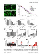
Figure 4. Diffusion behavior of nanoparticles in living cells and in vitro. (A–B’) Sample micrographs: HEp-2 cells were incubated with
fluorescent COOH-PS [YO] NPs for 30 min and FCS measurements were performed in the nucleus (A, yellow cross) or at cytoplasmic locations outside
the strongly labelled regions (B, yellow cross). Bar, 10 mm. A‘ and B‘ show the same subcellular locations after the FCS measurement. (C)
PLOS ONE | www.plosone.org 6 April 2013 | Volume 8 | Issue 4 | e62018