Page 327 - First Aid for the USMLE Step 1 2020, Thirtieth edition [MedicalBooksVN.com]_Neat
P. 327

CARDIOvASCuLAR  ``CARdIOvASCulAR—EMbRYOlOGY       CARDIOvASCuLAR  ``CARdIOvASCulAR—ANATOMY            SECTION III      283


                   ` `CARdIOvASCulAR—ANATOMY

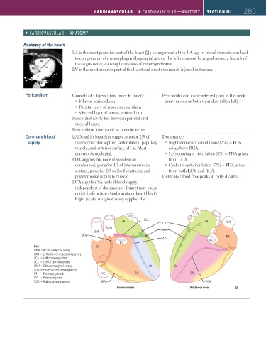
                  Anatomy of the heart
                   A                     LA is the most posterior part of the heart  A  ; enlargement of the LA (eg, in mitral stenosis) can lead
                                          to compression of the esophagus (dysphagia) and/or the left recurrent laryngeal nerve, a branch of
                          RV
                                          the vagus nerve, causing hoarseness (Ortner syndrome).
                      RA     LV          RV is the most anterior part of the heart and most commonly injured in trauma.

                        LA
                      pv
                          Ao
                   Pericardium           Consists of 3 layers (from outer to inner):  Pericarditis can cause referred pain to the neck,
                                             ƒ Fibrous pericardium                 arms, or one or both shoulders (often left).
                                             ƒ Parietal layer of serous pericardium
                                             ƒ Visceral layer of serous pericardium
                                         Pericardial cavity lies between parietal and
                                          visceral layers.
                                         Pericardium innervated by phrenic nerve.
                   Coronary blood        LAD and its branches supply anterior 2/3 of   Dominance:
                    supply                interventricular septum, anterolateral papillary     ƒ Right-dominant circulation (85%) = PDA
                                          muscle, and anterior surface of LV. Most   arises from RCA.
                                          commonly occluded.                          ƒ Left-dominant circulation (8%) = PDA arises
                                         PDA supplies AV node (dependent on          from LCX.
                                          dominance), posterior 1/3 of interventricular     ƒ Codominant circulation (7%) = PDA arises
                                          septum, posterior 2/3 walls of ventricles, and   from both LCX and RCA.
                                          posteromedial papillary muscle.         Coronary blood flow peaks in early diastole.
                                         RCA supplies SA node (blood supply
                                          independent of dominance). Infarct may cause
                                          nodal dysfunction (bradycardia or heart block).
                                          Right (acute) marginal artery supplies RV.


                                                                                                  PV
                                                                   PV
                                                                LA        LCA                         LA
                                                                                  LCX                           SVC
                                                        Aorta
                                                   SVC                            OMA
                                             RCA                                                                RA
                                                                                  LAD                   IVC
                                                               PT
                       Key:                         RA
                       AMA  = Acute marginal artery
                       LAD  = Left anterior descending artery
                       LCA  = Left coronary artery
                       LCX  = Left circumflex artery
                       OMA = Obtuse marginal artery                         LV          LV
                       PDA  = Posterior descending artery
                       PT     = Pulmonary trunk       IVC             RV                       RV
                       PV      = Pulmonary vein
                       RCA  = Right coronary artery   AMA                                             PDA
                                                             Anterior view                     Posterior view


          FAS1_2019_07-Cardio.indd   283                                                                                11/7/19   4:24 PM
   322   323   324   325   326   327   328   329   330   331   332