
Page 331 - First Aid for the USMLE Step 1 2020, Thirtieth edition [MedicalBooksVN.com]_Neat
P. 331
CARDIOvASCuLAR ``CARdIOvASCulAR—PHYSIOlOGY CARDIOvASCuLAR ``CARdIOvASCulAR—PHYSIOlOGY SECTION III 287
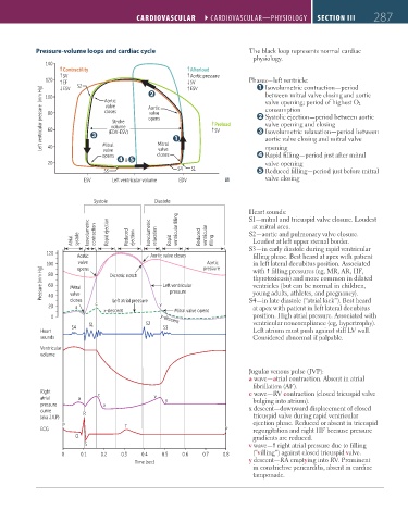
Pressure-volume loops and cardiac cycle The black loop represents normal cardiac
physiology.
140
Contractility Afterload
SV Aortic pressure
120 Phases—left ventricle:
EF S2 SV Isovolumetric contraction—period
ESV
ESV
Left ventricular pressure (mm Hg) 80 closes Aortic Preload Systolic ejection—period between aortic
between mitral valve closing and aortic
100
valve opening; period of highest O 2
Aortic
valve
consumption
valve
opens
Stroke
valve opening and closing
volume
60
Isovolumetric relaxation—period between
SV
(EDV-ESV)
aortic valve closing and mitral valve
Mitral
opening
40
valve
valve
closes
opens Mitral Rapid filling—period just after mitral
20 & valve opening
S3 S4 S1 Reduced filling—period just before mitral
ESV Left ventricular volume EDV valve closing
Systole Diastole
Heart sounds:
S1—mitral and tricuspid valve closure. Loudest
at mitral area.
Atrial systole Isovolumetric contraction Rapid ejection Reduced ejection Isovolumetric relaxation Rapid ventricular filling Reduced ventricular filling S2—aortic and pulmonary valve closure.
Loudest at left upper sternal border.
S3—in early diastole during rapid ventricular
120
Aortic Aortic valve closes pressure filling phase. Best heard at apex with patient
in left lateral decubitus position. Associated
valve
Aortic
Pressure (mm Hg) 100 Mitral Dicrotic notch Left ventricular with filling pressures (eg, MR, AR, HF,
opens
80
thyrotoxicosis) and more common in dilated
ventricles (but can be normal in children,
60
pressure
young adults, athletes, and pregnancy).
valve
40
20 closes c Left atrial pressure v S4—in late diastole (“atrial kick”). Best heard
at apex with patient in left lateral decubitus
a
x-descent Mitral valve opens
0 y-descent position. High atrial pressure. Associated with
S1 S2 ventricular noncompliance (eg, hypertrophy).
S4 S3
Heart Left atrium must push against stiff LV wall.
sounds Considered abnormal if palpable.
Ventricular
volume
Jugular venous pulse (JVP):
a wave—atrial contraction. Absent in atrial
fibrillation (AF).
Right c c wave—RV contraction (closed tricuspid valve
atrial a v y bulging into atrium).
pressure x x descent—downward displacement of closed
curve R
(aka J.V.P) tricuspid valve during rapid ventricular
P T ejection phase. Reduced or absent in tricuspid
ECG P regurgitation and right HF because pressure
Q gradients are reduced.
S v wave— right atrial pressure due to filling
0 0.1 0.2 0.3 0.4 0.5 0.6 0.7 0.8 (“villing”) against closed tricuspid valve.
Time (sec) y descent—RA emptying into RV. Prominent
in constrictive pericarditis, absent in cardiac
tamponade.
FAS1_2019_07-Cardio.indd 287 11/7/19 4:24 PM