Page 499 - First Aid for the USMLE Step 1 2020, Thirtieth edition [MedicalBooksVN.com]_Neat
P. 499

Musculoskeletal, skin, and connective tissue  ` anatomy and physiology  Musculoskeletal, skin, and connective tissue  ` anatomy and physiology  section iii  455


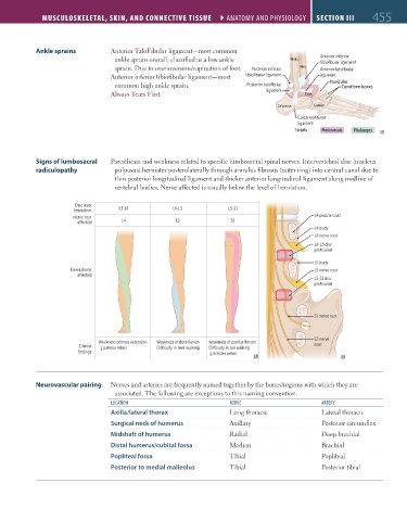
                  Ankle sprains          Anterior TaloFibular ligament—most common
                                          ankle sprain overall, classified as a low ankle      Fibula   Anterior inferior
                                                                                                        tibiofibular ligament
                                          sprain. Due to overinversion/supination of foot.   Posterior inferior  Tibia  Anterior talofibular
                                         Anterior inferior tibiofibular ligament—most   tibiofibular ligament  ligament
                                          common high ankle sprain.               Posterior talofibular     Navicular
                                                                                                              Cuneiform bones
                                         Always Tears First.                            ligament   Talus

                                                                                           Calcaneus  Cuboid
                                                                                                 Calcaneofibular
                                                                                                 ligament
                                                                                                Tarsals  Metatarsals  Phalanges


                  Signs of lumbosacral   Paresthesia and weakness related to specific lumbosacral spinal nerves. Intervertebral disc (nucleus
                  radiculopathy           pulposus) herniates posterolaterally through annulus fibrosus (outer ring) into central canal due to
                                          thin posterior longitudinal ligament and thicker anterior longitudinal ligament along midline of
                                          vertebral bodies. Nerve affected is usually below the level of herniation.


                              Disc level
                              herniation   L3-L4            L4-L5           L5-S1
                              Nerve root                                                              L4 pedicle (cut)
                                            L4              L5               S1
                                                                                                      L4 body
                                                                                                      L4 nerve root
                                                                                                      L4-L5 disc
                                                                                                      protrusion
                                                                                                      L5 body
                             Dermatome                                                                L5 nerve root
                                                                                                      L5-S1 disc
                                                                                                      protrusion


                                                                                                      S1 nerve root


                                                                                                      S2 nerve
                                     Weakness of knee extension  Weakness of dorsiflexion  Weakness of plantar flexion
                               Clinical       patellar reflex  Di€culty in heel walking  Di€culty in toe walking  root
                                      ↓
                               findings                                    Achilles reflex
                                                                       ↓


                  Neurovascular pairing  Nerves and arteries are frequently named together by the bones/regions with which they are
                                          associated. The following are exceptions to this naming convention.
                                         loCation                            nERVE                      aRtERy
                                         Axilla/lateral thorax               Long thoracic              Lateral thoracic
                                         Surgical neck of humerus            Axillary                   Posterior circumflex
                                         Midshaft of humerus                 Radial                     Deep brachial
                                         Distal humerus/cubital fossa        Median                     Brachial
                                         Popliteal fossa                     Tibial                     Popliteal
                                         Posterior to medial malleolus       Tibial                     Posterior tibial


          FAS1_2019_11-Musculo.indd   455                                                                               11/7/19   5:23 PM
   494   495   496   497   498   499   500   501   502   503   504