
Page 557 - First Aid for the USMLE Step 1 2020, Thirtieth edition [MedicalBooksVN.com]_Neat
P. 557
Neurology aNd Special SeNSeS ` neurology—PAthology Neurology aNd Special SeNSeS ` neurology—PAthology SecTioN iii 513
Intracranial hemorrhage
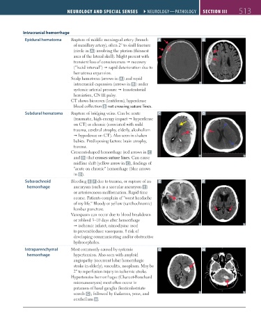
Epidural hematoma Rupture of middle meningeal artery (branch A B
of maxillary artery), often 2° to skull fracture
(circle in A ) involving the pterion (thinnest
area of the lateral skull). Might present with
transient loss of consciousness recovery
(“lucid interval”) rapid deterioration due to
hematoma expansion.
Scalp hematoma (arrows in A ) and rapid
intracranial expansion (arrows in B ) under
systemic arterial pressure transtentorial
herniation, CN III palsy.
CT shows biconvex (lentiform), hyperdense
blood collection B not crossing suture lines.
Subdural hematoma Rupture of bridging veins. Can be acute C D
(traumatic, high-energy impact hyperdense
on CT) or chronic (associated with mild
trauma, cerebral atrophy, elderly, alcoholism
hypodense on CT). Also seen in shaken
babies. Predisposing factors: brain atrophy,
trauma.
Crescent-shaped hemorrhage (red arrows in C
and D) that crosses suture lines. Can cause
midline shift (yellow arrow in C ), findings of
“acute on chronic” hemorrhage (blue arrows
in D).
Subarachnoid Bleeding E F due to trauma, or rupture of an E F
hemorrhage aneurysm (such as a saccular aneurysm E )
or arteriovenous malformation. Rapid time
course. Patients complain of “worst headache
of my life.” Bloody or yellow (xanthochromic)
lumbar puncture.
Vasospasm can occur due to blood breakdown
or rebleed 3–10 days after hemorrhage
ischemic infarct; nimodipine used
to prevent/reduce vasospasm. risk of
developing communicating and/or obstructive
hydrocephalus.
Intraparenchymal Most commonly caused by systemic G H
hemorrhage hypertension. Also seen with amyloid
angiopathy (recurrent lobar hemorrhagic
stroke in elderly), vasculitis, neoplasm. May be
2º to reperfusion injury in ischemic stroke.
Hypertensive hemorrhages (Charcot-Bouchard
microaneurysm) most often occur in
putamen of basal ganglia (lenticulostriate
vessels G), followed by thalamus, pons, and
cerebellum H.
FAS1_2019_12-Neurol.indd 513 11/8/19 7:39 AM