
Page 556 - First Aid for the USMLE Step 1 2020, Thirtieth edition [MedicalBooksVN.com]_Neat
P. 556
512 SecTioN iii Neurology aNd Special SeNSeS ` neurology—PAthology Neurology aNd Special SeNSeS ` neurology—PAthology
Ischemic brain Irreversible neuronal injury begins after 5 minutes of hypoxia. Most vulnerable: hippocampus,
disease/stroke neocortex, cerebellum (Purkinje cells), watershed areas (“vulnerable hippos need pure water”).
Stroke imaging: noncontrast CT to exclude hemorrhage (before tPA can be given). CT detects
ischemic changes in 6–24 hr. Diffusion-weighted MRI can detect ischemia within 3–30 min.
time sinCe isChemiC 12–24 hours 24–72 hours 3–5 dAys 1–2 WeeKs > 2 WeeKs
eVent
Histologic Eosinophilic Necrosis + Macrophages Reactive gliosis Glial scar
features cytoplasm neutrophils (microglia) (astrocytes)
+ pyknotic + vascular
nuclei (red proliferation
neurons)
Ischemic stroke Acute blockage of vessels disruption of blood flow and subsequent ischemia infarction
A liquefactive necrosis.
3 types:
Thrombotic—due to a clot forming directly at site of infarction (commonly the MCA A ),
usually over a ruptured atherosclerotic plaque.
Embolic—embolus from another part of the body obstructs vessel. Can affect multiple vascular
territories. Examples: atrial fibrillation, carotid artery stenosis, DVT with patent foramen ovale,
infective endocarditis.
Hypoxic—due to hypoperfusion or hypoxemia. Common during cardiovascular surgeries, tends
to affect watershed areas.
Treatment: tPA (if within 3–4.5 hr of onset and no hemorrhage/risk of hemorrhage) and/or
thrombectomy (if large artery occlusion). Reduce risk with medical therapy (eg, aspirin,
clopidogrel); optimum control of blood pressure, blood sugars, lipids; smoking cessation; and treat
conditions that risk (eg, atrial fibrillation, carotid artery stenosis).
Transient ischemic Brief, reversible episode of focal neurologic dysfunction without acute infarction (⊝ MRI), with the
attack majority resolving in < 15 minutes; ischemia (eg, embolus, small vessel stenosis).
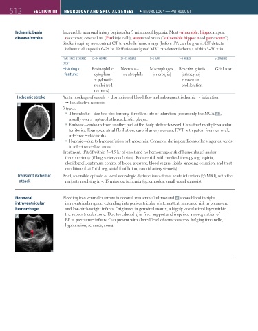
Neonatal Bleeding into ventricles (arrow in coronal transcranial ultrasound A shows blood in right
intraventricular intraventricular space, extending into periventricular white matter). Increased risk in premature
hemorrhage and low-birth-weight infants. Originates in germinal matrix, a highly vascularized layer within
A the subventricular zone. Due to reduced glial fiber support and impaired autoregulation of
BP in premature infants. Can present with altered level of consciousness, bulging fontanelle,
hypotension, seizures, coma.
FAS1_2019_12-Neurol.indd 512 11/8/19 7:39 AM