
Page 123 - Basic Principles of Textile Coloration
P. 123
112 PROTEIN FIBRES
water. Exactly the same types of interactions occur between different parts of the
same molecule, as between different molecules. These various types of interactions
are responsible for stabilising the particular configuration that a protein molecule
adopts and for many of its chemical and physical properties.
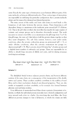
The ionic nature of the acidic and basic side-chains in wool leads to the
formation of salt links between the protein chains. Their formation is pH
dependent, being at a maximum at the isoelectric point around pH 5.5 (Scheme
7.1). This is the pH value at which the wool fibre has exactly the same number of
cationic and anionic groups and is therefore electrically neutral. The work
necessary to extend a wool fibre is at a maximum in the pH range from 5 to 9. In
this pH range, the ionic salt links help to hold the protein chains together so that
they resist elongation. The salt links cannot, however, exist under acidic
conditions, when the anionic carboxylate groups are protonated (pH < 5), or
under alkaline conditions, when the cationic ammonium ion groups are
deprotonated (pH > 9). Wool contains about 820 mmol kg–1 of amino groups and
a slightly lower number of carboxylic acid groups. These are responsible for its
ability to absorb large amounts of alkalis and acids, and for dyeing processes
involving ion exchange.
NH3 Wool CO2H H+ NH3 Wool CO2 H+ NH2 Wool CO2
acidic pH <5 isoelectric pH ~5.5 alkaline pH >9
Scheme 7.1
The disulphide bonds between adjacent protein chains, and between different
sections of the same chain, are a consequence of the incorporation of the double
amino acid cystine. These covalent crosslinks contribute to the stability of wool
fibres and to their mechanical, chemical and physical properties. There are also
amide or isopeptide covalent crosslinks, as for example that formed between
glutamic acid and lysine residues.
X-ray diffraction of unstretched wool fibres shows a pattern characteristic of a-
keratin, in which the individual protein molecules have a helical configuration and
wrap around each other in a helix. On stretching the wool fibre, the X-ray
diffraction pattern changes to that of b-keratin, in which the chains are fully